

Functional and structural characterization of thermostable D-amino acid aminotransferases from Geobacillus spp
- PMID: 16461714
- PMCID: PMC1392904
- DOI: 10.1128/AEM.72.2.1588-1594.2006
Functional and structural characterization of thermostable D-amino acid aminotransferases from Geobacillus spp
Abstract
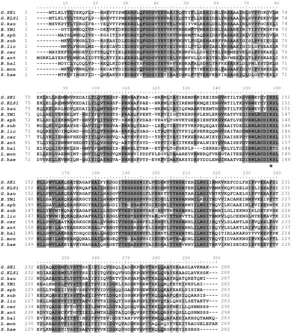
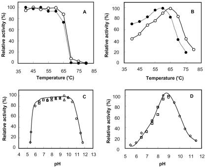
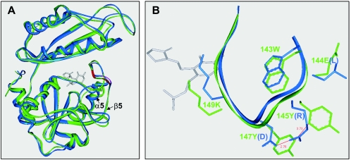
D-amino acid aminotransferases (D-AATs) from Geobacillus toebii SK1 and Geobacillus sp. strain KLS1 were cloned and characterized from a genetic, catalytic, and structural aspect. Although the enzymes were highly thermostable, their catalytic capability was approximately one-third of that of highly active Bacilli enzymes, with respective turnover rates of 47 and 55 s(-1) at 50 degrees C. The Geobacillus enzymes were unique and shared limited sequence identities of below 45% with D-AATs from mesophilic and thermophilic Bacillus spp., except for a hypothetical protein with a 72% identity from the G. kaustophilus genome. Structural alignments showed that most key residues were conserved in the Geobacillus enzymes, although the conservative residues just before the catalytic lysine were distinctively changed: the 140-LRcD-143 sequence in Bacillus D-AATs was 144-EYcY-147 in the Geobacillus D-AATs. When the EYcY sequence from the SK1 enzyme was mutated into LRcD, a 68% increase in catalytic activity was observed, while the binding affinity toward alpha-ketoglutarate decreased by half. The mutant was very close to the wild-type in thermal stability, indicating that the mutations did not disturb the overall structure of the enzyme. Homology modeling also suggested that the two tyrosine residues in the EYcY sequence from the Geobacillus D-AATs had a pi/pi interaction that was replaceable with the salt bridge interaction between the arginine and aspartate residues in the LRcD sequence.
Figures
Similar articles
-
Characterization of thermostable lipase from thermophilic Geobacillus sp. TW1.Protein Expr Purif. 2005 Jul;42(1):153-9. doi: 10.1016/j.pep.2005.03.011. Epub 2005 Mar 31. Protein Expr Purif. 2005. PMID: 15939301
-
Gene cloning, expression, and crystallization of a thermostable exo-inulinase from Geobacillus stearothermophilus KP1289.Appl Microbiol Biotechnol. 2003 Aug;62(2-3):180-5. doi: 10.1007/s00253-003-1261-3. Epub 2003 Feb 22. Appl Microbiol Biotechnol. 2003. PMID: 12883863
-
Characterization of a novel thermostable carboxylesterase from Geobacillus kaustophilus HTA426 shows the existence of a new carboxylesterase family.J Bacteriol. 2009 May;191(9):3076-85. doi: 10.1128/JB.01060-08. Epub 2009 Mar 20. J Bacteriol. 2009. PMID: 19304850 Free PMC article.
-
Peculiarities and biotechnological potential of environmental adaptation by Geobacillus species.Appl Microbiol Biotechnol. 2018 Dec;102(24):10425-10437. doi: 10.1007/s00253-018-9422-6. Epub 2018 Oct 11. Appl Microbiol Biotechnol. 2018. PMID: 30310966 Review.
-
Leucine Dehydrogenase: Structure and Thermostability.Subcell Biochem. 2021;96:355-372. doi: 10.1007/978-3-030-58971-4_10. Subcell Biochem. 2021. PMID: 33252736 Review.
Cited by
-
d-Amino Acids and Lactic Acid Bacteria.Microorganisms. 2019 Dec 12;7(12):690. doi: 10.3390/microorganisms7120690. Microorganisms. 2019. PMID: 31842512 Free PMC article. Review.
-
Diversity of D-Amino Acid Utilizing Bacteria From Kongsfjorden, Arctic and the Metabolic Pathways for Seven D-Amino Acids.Front Microbiol. 2020 Jan 10;10:2983. doi: 10.3389/fmicb.2019.02983. eCollection 2019. Front Microbiol. 2020. PMID: 31998270 Free PMC article.
-
Enantioselective Utilization of D-Amino Acids by Deep-Sea Microorganisms.Front Microbiol. 2016 Apr 19;7:511. doi: 10.3389/fmicb.2016.00511. eCollection 2016. Front Microbiol. 2016. PMID: 27148200 Free PMC article.
-
Improvement in catalytic activity and thermostability of a GH10 xylanase and its synergistic degradation of biomass with cellulase.Biotechnol Biofuels. 2019 Dec 3;12:278. doi: 10.1186/s13068-019-1620-7. eCollection 2019. Biotechnol Biofuels. 2019. PMID: 31827606 Free PMC article.
-
Assessment of Metabolic Changes in Mycobacterium smegmatis Wild-Type and alr Mutant Strains: Evidence of a New Pathway of d-Alanine Biosynthesis.J Proteome Res. 2017 Mar 3;16(3):1270-1279. doi: 10.1021/acs.jproteome.6b00871. Epub 2017 Feb 14. J Proteome Res. 2017. PMID: 28121156 Free PMC article.
References
-
- Ager, D. J., I. G. Fotheringham, S. A. Laneman, D. P. Pantaleone, and P. P. Taylor. 2000. The large scale synthesis of “unnatural” amino acids. Enantiomer 5:235-243. - PubMed
-
- Ash, C., J. A. E. Farrow, S. Wallbanks, and M. D. Collins. 1991. Phylogenetic heterogeneity of the genus bacillus revealed by comparative analysis of small subunit rRNA sequences. Lett. Appl. Microbiol. 13:202-206.
-
- Bae, H.-S., S.-P. Hong, S.-G. Lee, M.-S. Kwak, N. Esaki, and M.-H. Sung. 2002. Application of a thermostable glutamate racemase from Bacillus sp. SK-1 for the production of d-phenylalanine in a multi-enzyme. Syst. J. Mol. Catal. B. Enzymatic 17:223-233.
-
- Berntsson, S. 1995. Spectrometric determination of pyruvic acid by the salicylaldehyde method. Anal. Biochem. 27:1659-1660.
Publication types
MeSH terms
Substances
LinkOut - more resources
Full Text Sources
Other Literature Sources
Research Materials
Miscellaneous
