

Txp40, a ubiquitous insecticidal toxin protein from Xenorhabdus and Photorhabdus bacteria
- PMID: 16461722
- PMCID: PMC1392922
- DOI: 10.1128/AEM.72.2.1653-1662.2006
Txp40, a ubiquitous insecticidal toxin protein from Xenorhabdus and Photorhabdus bacteria
Abstract
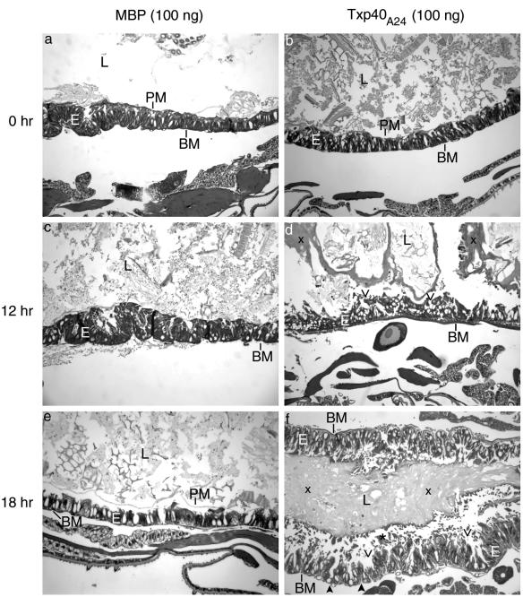
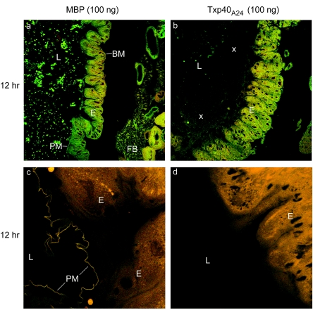
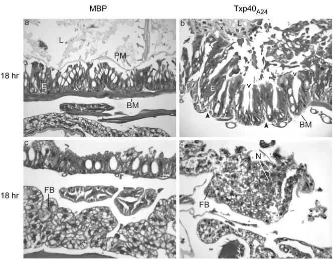
Xenorhabdus and Photorhabdus are gram-negative bacteria that produce a range of proteins that are toxic to insects. We recently identified a novel 42-kDa protein from Xenorhabdus nematophila that was lethal to the larvae of insects such as Galleria mellonella and Helicoverpa armigera when it was injected at doses of 30 to 40 ng/g larvae. In the present work, the toxin gene txp40 was identified in another 59 strains of Xenorhabdus and Photorhabdus, indicating that it is both highly conserved and widespread among these bacteria. Recombinant toxin protein was shown to be active against a variety of insect species by direct injection into the larvae of the lepidopteran species G. mellonella, H. armigera, and Plodia interpunctella and the dipteran species Lucilia cuprina. The protein exhibited significant cytotoxicity against two dipteran cell lines and two lepidopteran cell lines but not against a mammalian cell line. Histological data from H. armigera larvae into which the toxin was injected suggested that the primary site of action of the toxin is the midgut, although some damage to the fat body was also observed.
Figures
Similar articles
-
PirAB protein from Xenorhabdus nematophila HB310 exhibits a binary toxin with insecticidal activity and cytotoxicity in Galleria mellonella.J Invertebr Pathol. 2017 Sep;148:43-50. doi: 10.1016/j.jip.2017.04.007. Epub 2017 Apr 21. J Invertebr Pathol. 2017. PMID: 28438456
-
Txp40, an insecticidal toxin protein from Xenorhabdus nematophila: Purification, toxicity assessment and biophysical characterization.Toxicon. 2022 Oct 30;218:40-46. doi: 10.1016/j.toxicon.2022.09.003. Epub 2022 Sep 9. Toxicon. 2022. PMID: 36096207
-
Txp40, a protein from Photorhabdus akhurstii, conferred potent insecticidal activity against the larvae of Helicoverpa armigera, Spodoptera litura and S. exigua.Pest Manag Sci. 2020 Jun;76(6):2004-2014. doi: 10.1002/ps.5732. Epub 2020 Jan 8. Pest Manag Sci. 2020. PMID: 31867818
-
The great potential of entomopathogenic bacteria Xenorhabdus and Photorhabdus for mosquito control: a review.Parasit Vectors. 2020 Jul 29;13(1):376. doi: 10.1186/s13071-020-04236-6. Parasit Vectors. 2020. PMID: 32727530 Free PMC article. Review.
-
Mutualism and pathogenesis in Xenorhabdus and Photorhabdus: two roads to the same destination.Mol Microbiol. 2007 Apr;64(2):260-8. doi: 10.1111/j.1365-2958.2007.05671.x. Mol Microbiol. 2007. PMID: 17493120 Review.
Cited by
-
Type VI secretion system MIX-effectors carry both antibacterial and anti-eukaryotic activities.EMBO Rep. 2017 Nov;18(11):1978-1990. doi: 10.15252/embr.201744226. Epub 2017 Sep 14. EMBO Rep. 2017. PMID: 28912123 Free PMC article.
-
A novel tumor-targeting strain of Xenorhabdus stockiae exhibits potent biological activities.Front Bioeng Biotechnol. 2022 Sep 7;10:984197. doi: 10.3389/fbioe.2022.984197. eCollection 2022. Front Bioeng Biotechnol. 2022. PMID: 36159678 Free PMC article.
-
Genome Sequence Analysis of Native Xenorhabdus Strains Isolated from Entomopathogenic Nematodes in Argentina.Toxins (Basel). 2024 Feb 17;16(2):108. doi: 10.3390/toxins16020108. Toxins (Basel). 2024. PMID: 38393187 Free PMC article.
-
Attenuated virulence and genomic reductive evolution in the entomopathogenic bacterial symbiont species, Xenorhabdus poinarii.Genome Biol Evol. 2014 Jun 5;6(6):1495-513. doi: 10.1093/gbe/evu119. Genome Biol Evol. 2014. PMID: 24904010 Free PMC article.
-
Variations of Indole Metabolites and NRPS-PKS Loci in Two Different Virulent Strains of Xenorhabdus hominickii.Front Microbiol. 2020 Nov 24;11:583594. doi: 10.3389/fmicb.2020.583594. eCollection 2020. Front Microbiol. 2020. PMID: 33329448 Free PMC article.
References
-
- Akhurst, R. J., and N. E. Boemare. 1988. A numerical taxonomic study of the genus Xenorhabdus (Enterobacteriaceae) and proposed elevation of the subspecies of X. nematophilus to species. J. Gen. Microbiol. 134:1835-1845. - PubMed
-
- Akhurst, R. J., and G. B. Dunphy. 1993. Tripartite interactions between symbiotically associated entomopathogenic bacteria, nematodes, and their insect hosts, p. 1-23. In N. E. Beckage, S. N. Thompson, and B. A. Federici (ed.), Parasites and pathogens of insects, vol. 2. Academic Press, Inc., San Diego, Calif.
-
- Akhurst, R. J., A. J. Smigielski, J. Mari, N. Boemare, and R. G. Mourant. 1992. Restriction analysis of phase variation in Xenorhabdus spp. (Enterobacteriaceae), entomopathogenic bacteria associated with nematodes. Syst. Appl. Microbiol. 15:469-473.
-
- Au, C., P. Dean, S. E. Reynolds, and R. H. ffrench-Constant. 2004. Effect of the insect pathogenic bacterium Photorhabdus on insect phagocytes. Cell. Microbiol. 6:89-95. - PubMed
MeSH terms
Substances
Associated data
- Actions
- Actions
- Actions
- Actions
- Actions
- Actions
- Actions
- Actions
- Actions
- Actions
- Actions
- Actions
LinkOut - more resources
Full Text Sources
Other Literature Sources
Molecular Biology Databases
