. 2006 Feb 14;103(7):2052-6.
doi: 10.1073/pnas.0511078103. Epub 2006 Feb 6.

Fast complementation of split fluorescent protein triggered by DNA hybridization

Affiliations

Fast complementation of split fluorescent protein triggered by DNA hybridization

Vadim V Demidov et al. Proc Natl Acad Sci U S A. .

Abstract

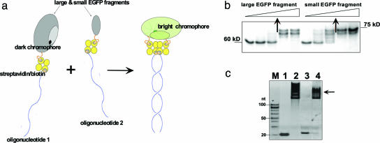
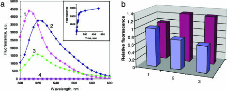
Fluorescent proteins have proven to be excellent reporters and biochemical sensors with a wide range of applications. In a split form, they are not fluorescent, but their fluorescence can be restored by supplementary protein-protein or protein-nucleic acid interactions that reassemble the split polypeptides. However, in prior studies, it took hours to restore the fluorescence of a split fluorescent protein because the formation of the protein chromophore slowly occurred de novo concurrently with reassembly. Here we provide evidence that a fluorogenic chromophore can self-catalytically form within an isolated N-terminal fragment of the enhanced green fluorescent protein (EGFP). We show that restoration of the split protein fluorescence can be driven by nucleic acid complementary interactions. In our assay, fluorescence development is fast (within a few minutes) when complementary oligonucleotide-linked fragments of the split EGFP are combined. The ability of our EGFP system to respond quickly to DNA hybridization should be useful for detecting the kinetics of many other types of pairwise interactions both in vitro and in living cells.


Conflict of interest statement

Conflict of interest statement: No conflicts declared.

Figures

Fig. 1.
Fig. 1.
Structure of the large EGFP fragment (1–158 N-terminal amino acids) analyzed by DMD simulations. (a) Backbone representation of 10 folded and aligned structures of the large EGFP fragment obtained in DMD simulations at T = 0.3 (T is measured in ε/KB units). The segment from 62 to 70 amino acids, containing the chromophore-forming amino acids (T66, Y67, and G68), is colored blue. The C terminus of this polypeptide is very flexible because of a small number of contacts with the rest of the molecule, so the alignment was made by omitting these amino acids. (b) The root-mean-square deviation (RMSD) of each residue in the folded large EGFP fragment relative to the intact EGFP structure as a function of temperature. The chromophore-forming residues are in the shaded region, and their spatial arrangement at lower temperatures is essentially fixed, with deviation ≤2Å.
Fig. 2.
Fig. 2.
Characteristics of EGFP fragments overexpressed in E. coli and isolated by using the intein self-splicing technology. (a) Fifteen percent SDS/PAGE analysis of protein samples containing the large (lanes 1) and small (lanes 2) EGFP fragments (two samples of each fragment from different protein preparations are shown as examples). Lane M corresponds to a molecular mass protein ladder. Large and small EGFP fragments are seen as ≈15 kDa and ≈10 kDa bands, respectively (marked with asterisks). Although the small EGFP fragment is practically pure, the large EGFP fragment is somewhat contaminated by intein (≈25 kDa) and unsplit fusion (≈40 kDa). (b) Fluorescence excitation (curve 1) and fluorescence emission (curve 2) spectra of the large EGFP fragment (2 μM in PBS buffer, pH 7.4).
Fig. 3.
Fig. 3.
Design and assembly of a nucleic acid-supported EGFP complementation system with rapid signal response. (a) Schematics of the complementation of split fluorescent protein by DNA hybridization. Fluorescent protein (EGFP) is dissected into two nonfluorescent fragments, one of which contains preformed chromophore capable of bright fluorescence within a full-size protein. Both protein fragments are linked to complementary oligonucleotides via biotin–streptavidin interactions. In our protocol, we endeavor to create a 1:1:1 ratio of protein/streptavidin/oligonucleotide complex. In a mixture, the two nucleoprotein constructs associate by sequence-specific duplex DNA formation, which triggers complementation of the large and small EGFP fragments, resulting in fast development of fluorescence. (b) Gel-shift assay (10% SDS/PAGE) showing binding of increased amounts of biotinylated EGFP fragments with a fixed amount of streptavidin (2 μg; 60-kDa band). Arrows indicate the protein amounts resulting in 1:1 complexes (70- to 75-kDa bands), which correspond to ≥70% yield of biotinylation. (c) Gel-shift assay (10% PAGE) demonstrating the formation of 1:1:1 tripartite molecular constructions depicted in Fig. 1a and comprising the large or small EGFP fragment, streptavidin, and a corresponding oligonucleotide (see Table 1, which is published as supporting information on the PNAS web site, for their sequences). Lanes 1 and 2, biotinylated oligo 1 in the absence (1) or presence (2) of the large EGFP fragment coupled to streptavidin; lanes 3 and 4, biotinylated oligonucleotide 2 in the absence (3) or presence (4) of the small EGFP fragment coupled to streptavidin; M, 20-bp size marker. Arrow marks the position of the required oligonucleotide–protein complexes that are strongly shifted upward as expected.
Fig. 4.
Fig. 4.
Fluorescent responses of the split EGFP system upon DNA hybridization. (a) Fluorescence spectra of intact EGFP (1) and of the split EGFP-based protein complex reassembled by DNA hybridization from the tripartite molecular constructions (2), each taken at ≈200 nM concentrations in PBS buffer at pH 7.4 (spectra recorded 20 min after mixing) (3), the same as sample 2 plus 100-fold excess of one of the two complementary oligonucleotides (nonbiotinylated oligo 1) (4), and control containing both EGFP fragments coupled to streptavidin but without oligonucleotides. (Inset) The time course of the fluorescence development in sample 2 was recorded at 524 nm. (b) Effect of Mg2+ cations on intact EGFP (blue) and on the reassembled split EGFP complex containing duplex DNA (purple). Column 1, no Mg2+; columns 2 and 3: 2 min and 3 h, respectively, after addition of 2 mM Mg2+.

References

    1. Ozawa T., Sako Y., Sato M., Kitamura T., Umezawa Y. Nat. Biotechnol. 2003;21:287–293. - PubMed
    1. Hu C. D., Kerppola T. K. Nat. Biotechnol. 2003;21:539–545. - PMC - PubMed
    1. Remy I., Michnick S. W. Methods. 2004;32:381–388. - PubMed
    1. Magliery T. J., Wilson C. G., Pan W., Mishler D., Ghosh I., Hamilton A. D., Regan L. J. Am. Chem. Soc. 2005;127:146–157. - PubMed
    1. Stains C. I., Porter J. R., Ooi A. T., Segal D. J., Ghosh I. J. Am. Chem. Soc. 2005;127:10782–10783. - PubMed

Publication types

MeSH terms

LinkOut - more resources