. 2006 Feb 14;103(7):2098-102.
doi: 10.1073/pnas.0511026103. Epub 2006 Feb 6.

Lateral mobility of proteins in liquid membranes revisited

Affiliations

Lateral mobility of proteins in liquid membranes revisited

Y Gambin et al. Proc Natl Acad Sci U S A. .

Abstract

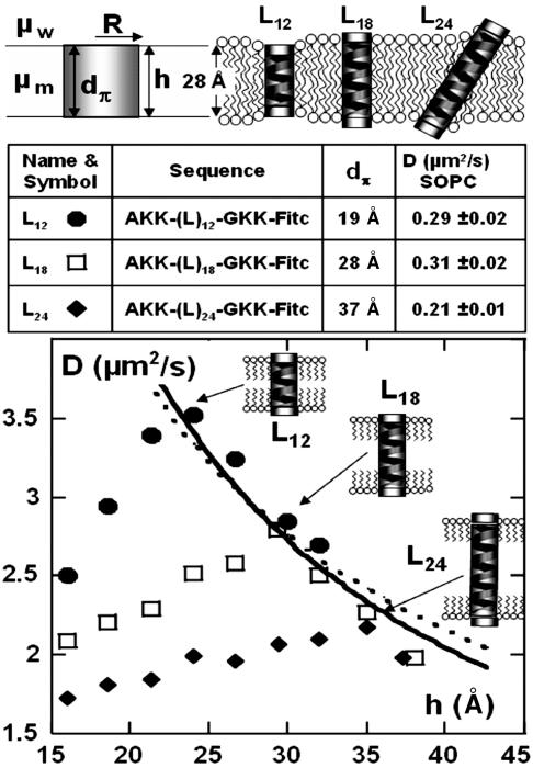
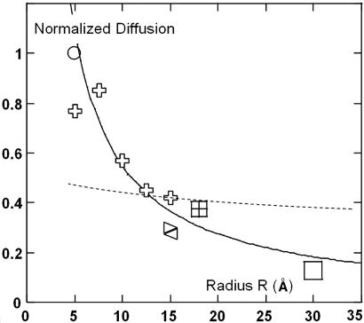
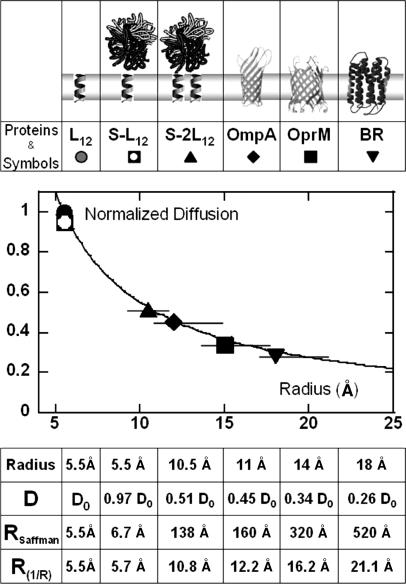
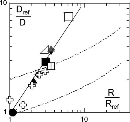
The biological function of transmembrane proteins is closely related to their insertion, which has most often been studied through their lateral mobility. For >30 years, it has been thought that hardly any information on the size of the diffusing object can be extracted from such experiments. Indeed, the hydrodynamic model developed by Saffman and Delbrück predicts a weak, logarithmic dependence of the diffusion coefficient D with the radius R of the protein. Despite widespread use, its validity has never been thoroughly investigated. To check this model, we measured the diffusion coefficients of various peptides and transmembrane proteins, incorporated into giant unilamellar vesicles of 1-stearoyl-2-oleoyl-sn-glycero-3-phosphocholine (SOPC) or in model bilayers of tunable thickness. We show in this work that, for several integral proteins spanning a large range of sizes, the diffusion coefficient is strongly linked to the protein dimensions. A heuristic model results in a Stokes-like expression for D, (D proportional, variant 1/R), which fits literature data as well as ours. Diffusion measurement is then a fast and fruitful method; it allows determining the oligomerization degree of proteins or studying lipid-protein and protein-protein interactions within bilayers.


Conflict of interest statement

Conflict of interest statement: No conflicts declared.

Figures

Fig. 1.
Fig. 1.
Peptides used and their D variations versus bilayer thickness h. (Top) The parameters used in the Saffman–Delbrück model (Eq. 1), in the case of peptides diffusing in a giant unilamellar vesicle (GUV) made of SOPC. (Middle) A summary of the properties of the peptides used, their sequences, and their hydrophobic length dπ. The right-hand column gives the measured diffusion coefficient of these peptides in single GUVs. The diffusion coefficient was determined by using evanescent fluorescence recovery after pattern photobleaching technique. The data are typically averaged >200 vesicles. (Bottom) The variation of the diffusion due to the swelling of the C12E5 bilayer for the three analog peptides L12 (•), L18 (□), and L24 (♦). For each peptide, five sets of experiments allowed us to obtain average values with a reproducibility of >5% (the symbol size). The dotted line is the fit with the Saffman–Delbrück model (Eq. 1), using only one adjustable parameter, μm. The radius was taken as 5.5 Å and the viscosity of water as 0.01 P, leading to μm = 2.94 P. The solid line represents a simple 1/h dependence. Note that the relative D variations are the same in C12E5 bilayers and in SOPC membranes: for h = 28 Å, L24 diffuses 30% slower than L18; L12 and L18 have similar mobilities.
Fig. 2.
Fig. 2.
Normalized diffusion coefficient (D/Dlipid) vs. peptide radius R in lipid bilayers. Crosses correspond respectively from the left to monomers, dimers, trimers, tetramers, and hexamers of transmembrane peptides (15). The square symbols at R = 15, 18, and 30 Å correspond, respectively, to acetylcholine receptor (AChR), BR, and SR-ATPase (16). The solid line is a 1/R fit, and the dashed line represents the prediction of Saffman’s model, using h = 28 Å, μm = 1.75 P, and μw = 1 cP as in ref 15.
Fig. 3.
Fig. 3.
Normalized diffusion coefficient (D/DL12) vs. peptide radius R, in C12E5 bilayers. The formation of streptavidin-peptide assemblies is described in Materials and Methods. To avoid possible denaturation, the transmembrane proteins are embedded in dodecane-free membranes. The hydrophobic mismatch between protein height and bilayer thickness creates a local deformation. As discussed in Materials and Methods, this effect leads to a large uncertainty in the effective radius of the protein, represented by the horizontal bars in the plot. The diffusion coefficients are normalized by the diffusion of the L12 peptide (RL12 = 5.5 Å ≈ RSOPC), extrapolated to the thickness of a dry bilayer in Fig. 1 (D0 = 4.8 ± 0.2 μm2/s). From the measured D values and Eq. 1, one can estimate the corresponding where h = 16 Å, and μm = 2.94 P, μw = 10−2 P, as in Fig. 1. The dashed line is the fit using these parameters. For comparison, we indicate the radii calculated from the 1/R law (solid line, as in Fig. 2):
Fig. 4.
Fig. 4.
Normalized inverse diffusion coefficient Dref/D vs. object radius R/Rref, (open symbols are data gathered from the literature, and filled symbols are from this work). For peptide assemblies and proteins in C12E5 bilayers (filled symbols as in Fig. 3), the peptide L12 serves as reference: Dref/D = DL12/D; the BR in SOPC (gray triangle) is compared with the L18 peptide: Dref/D = DL18/D. As in Fig. 2, for oligomers of peptides (crosses), acetylcholine receptor (AChR), BR, and SR-ATPase (squares), the lipid diffusion serves as reference. The solid line is a power-law regression leading to Dref/DR1.04; for comparison, the dashed line represents the prediction of the Saffman–Delbrück model (Eq. 1) (upper line, same as in Fig. 3, and lower fit as in Fig. 2).

References

    1. Saffman P. G., Delbrück M. Proc. Natl. Acad. Sci. USA. 1975;72:3111–3113. - PMC - PubMed
    1. Reits E. A. J., Neefjes J. J. Nat. Cell Biol. 2001;3:145–147. - PubMed
    1. Kahya N., Pecheur E.-I., de Boeij W. P., Wiersam D. A., Hoekstra D. Biophys. J. 2001;81:1464–1474. - PMC - PubMed
    1. Tsapis N., Reiss-Husson F., Ober R., Genest M., Hodges R. S., Urbach W. Biophys. J. 2001;81:1613–1623. - PMC - PubMed
    1. Kenworthy A. K., Nichols B. J., Remmert C. L., Hendrix G. M., Kumar M., Zimmerberg J., Lippincott-Schwartz J. J. Cell Biol. 2004;165:735–746. - PMC - PubMed

Publication types

MeSH terms

Substances