

Screening for transglutaminase-catalyzed modifications by peptide mass finger printing using multipoint recalibration on recognized peaks for high mass accuracy
- PMID: 16461943
- PMCID: PMC2291725
Screening for transglutaminase-catalyzed modifications by peptide mass finger printing using multipoint recalibration on recognized peaks for high mass accuracy
Abstract
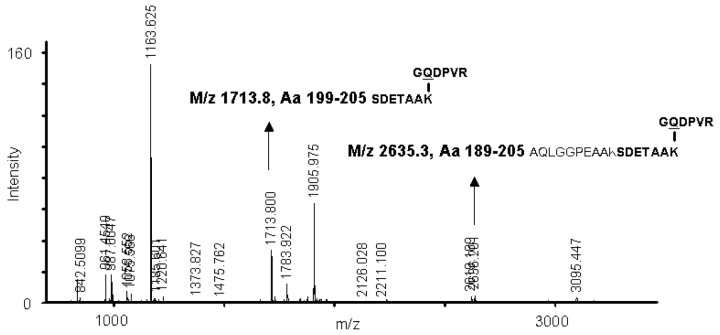
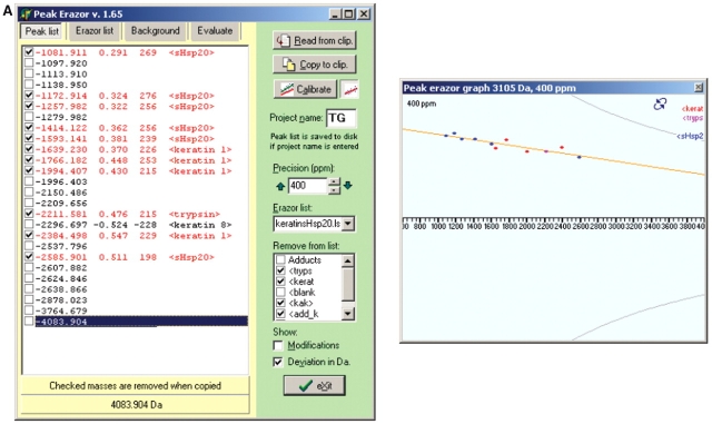
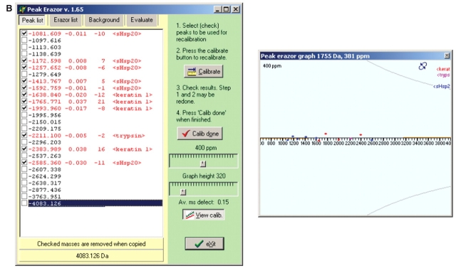
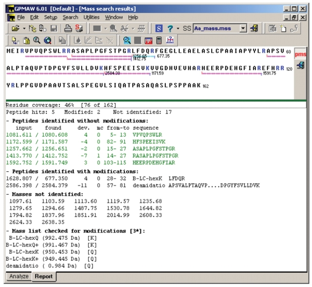
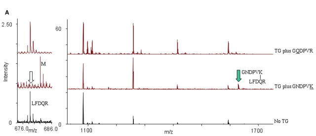
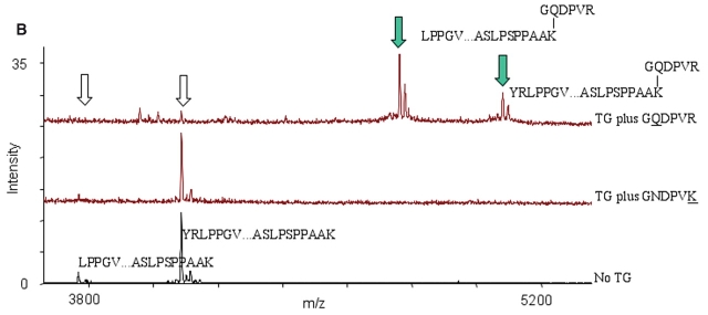
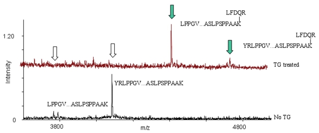
Detection of posttranslational modifications is expected to be one of the major future experimental challenges for proteomics. We describe herein a mass spectrometric procedure to screen for protein modifications by peptide mass fingerprinting that is based on post-data acquisition improvement of the mass accuracy by exporting the peptide mass values into analytical software for multipoint recalibration on recognized peaks. Subsequently, the calibrated peak mass data set is used in searching for modified peptides, i.e., peptides possessing specific mass deviations. In order to identify the location of Lys- and Gln-residues available for transglutaminase-catalyzed isopeptide bond formation, mammalian small heat shock proteins (sHsps) were screened for labeling with the two hexapeptide probes GQDPVR and GNDPVK in presence of transglutaminase. Peptide modification due to cross-linking of the GQDPVR hexa-peptide probe was detected for C-terminal Lys residues. Novel transglutaminase-susceptible Gln sites were identified in two sHsps (Q31/Q27 in Hsp20 and HspB2, respectively), by cross-linking of the GNDPVK hexapeptide probe. Deamidation of specific Gln residues was also detected, as well an isopeptide derived from intramolecular Gln-Lys isopeptide bond formation. We conclude that peptide mass fingerprinting can be an efficient way of screening for various posttranslational modifications. Basically any instrumentation for MALDI mass spectrometry can be used, provided that post-data acquisition recalibration is applied.
Figures
Similar articles
-
Site-specific transamidation and deamidation of the small heat-shock protein Hsp20 by tissue transglutaminase.Proteins. 2006 Mar 1;62(4):1044-52. doi: 10.1002/prot.20837. Proteins. 2006. PMID: 16385579
-
Transglutaminase 2-Catalyzed Intramolecular Cross-Linking of Osteopontin.Biochemistry. 2016 Jan 19;55(2):294-303. doi: 10.1021/acs.biochem.5b01153. Epub 2016 Jan 4. Biochemistry. 2016. PMID: 26678563
-
Mass spectrometric identification of formaldehyde-induced peptide modifications under in vivo protein cross-linking conditions.Anal Chim Acta. 2008 Jun 23;618(2):168-83. doi: 10.1016/j.aca.2008.04.049. Epub 2008 May 2. Anal Chim Acta. 2008. PMID: 18513538
-
Matrix-assisted laser desorption ionization mass spectrometry: applications in peptide and protein characterization.Protein Expr Purif. 1995 Apr;6(2):109-23. doi: 10.1006/prep.1995.1014. Protein Expr Purif. 1995. PMID: 7606158 Review.
-
GKVLAK–(Gadolinium-(1,4,7,10-tetraazacyclododecane-1,4,7-tris(acetic acid t-butyl ester)-10-acetic acid monoamide))–GGGGTVQQEL.2011 Mar 29 [updated 2011 Apr 26]. In: Molecular Imaging and Contrast Agent Database (MICAD) [Internet]. Bethesda (MD): National Center for Biotechnology Information (US); 2004–2013. 2011 Mar 29 [updated 2011 Apr 26]. In: Molecular Imaging and Contrast Agent Database (MICAD) [Internet]. Bethesda (MD): National Center for Biotechnology Information (US); 2004–2013. PMID: 21542560 Free Books & Documents. Review.
Cited by
-
Dissecting the mechanisms of tissue transglutaminase-induced cross-linking of alpha-synuclein: implications for the pathogenesis of Parkinson disease.J Biol Chem. 2009 May 8;284(19):13128-42. doi: 10.1074/jbc.M809067200. Epub 2009 Jan 21. J Biol Chem. 2009. PMID: 19164286 Free PMC article.
References
-
- Pandey A, Mann M. Proteomics to study genes and genomes. Nature, 2000;405: 837–846. - PubMed
-
- Larsen MR, Roepstorff P. Mass spectrometric identification of proteins and characterization of their posttranslational modifications in proteome analysis. Fresenius J Anal Chem, 2000;366:677–690. - PubMed
-
- Mann M, Jensen ON. Proteomic analysis of posttranslational modifications. Nat Biotechnol, 2003;21:255–261. - PubMed
-
- Jensen ON. Modification-specific proteomics: Characterization of posttranslational modifications by mass spectrometry. Curr Opin Chem Biol, 2004; 8: 33–41. - PubMed
-
- Khidekel N, Hsieh-Wilson LC. A “molecular switchboard”—Covalent modifications to proteins and their impact on transcription. Org Biomol Chem, 2004;2:1–7. - PubMed
Publication types
MeSH terms
Substances
LinkOut - more resources
Full Text Sources
Other Literature Sources