. 2006 Feb;2(2):e12.
doi: 10.1371/journal.pgen.0020012. Epub 2006 Feb 3.

A link between meiotic prophase progression and crossover control

Affiliations

A link between meiotic prophase progression and crossover control

Peter M Carlton et al. PLoS Genet. 2006 Feb.

Abstract

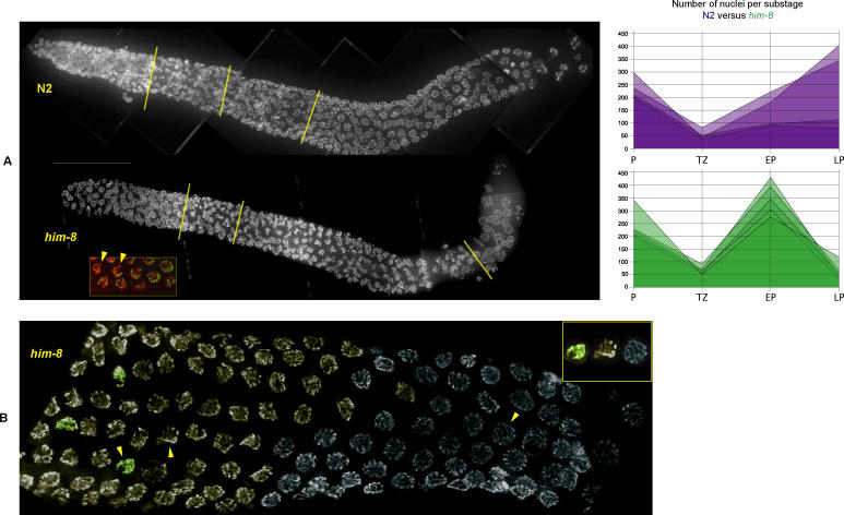
During meiosis, most organisms ensure that homologous chromosomes undergo at least one exchange of DNA, or crossover, to link chromosomes together and accomplish proper segregation. How each chromosome receives a minimum of one crossover is unknown. During early meiosis in Caenorhabditis elegans and many other species, chromosomes adopt a polarized organization within the nucleus, which normally disappears upon completion of homolog synapsis. Mutations that impair synapsis even between a single pair of chromosomes in C. elegans delay this nuclear reorganization. We quantified this delay by developing a classification scheme for discrete stages of meiosis. Immunofluorescence localization of RAD-51 protein revealed that delayed meiotic cells also contained persistent recombination intermediates. Through genetic analysis, we found that this cytological delay in meiotic progression requires double-strand breaks and the function of the crossover-promoting heteroduplex HIM-14 (Msh4) and MSH-5. Failure of X chromosome synapsis also resulted in impaired crossover control on autosomes, which may result from greater numbers and persistence of recombination intermediates in the delayed nuclei. We conclude that maturation of recombination events on chromosomes promotes meiotic progression, and is coupled to the regulation of crossover number and placement. Our results have broad implications for the interpretation of meiotic mutants, as we have shown that asynapsis of a single chromosome pair can exert global effects on meiotic progression and recombination frequency.


Conflict of interest statement

Author contributions. PMC and AFD conceived and designed the experiments. PMC and APF performed the experiments. PMC, APF, and AFD analyzed the data. PMC and AFD wrote the paper.

Figures

Figure 1
Figure 1. Criteria for Classifying Meiotic Nuclei into Substages of Prophase I
(A) Nuclei with even distribution of chromatin that were located distal to transition zone nuclei were classified as premeiotic. (B) Nuclei with asymmetric distribution of chromatin that could not be resolved into individual chromosomes were classified as transition zone. (C) Nuclei with asymmetric distribution of individually resolvable chromosomes were classified as early pachytene. (D) Nuclei with fully paired chromosomes distributed around the entire nucleus were classified as late pachytene. DNA (blue) was stained with DAPI; nucleoli (red) were stained with antibodies against nucleolar protein NOP-1.
Figure 2
Figure 2. Progression from Early to Late Pachytene Is Delayed in X Asynapsis Mutants
(A) Gonads imaged at 100× and composited. Top: N2; bottom: him-8(mn253). The three yellow lines demarcate a rough separation of the gonad into four sections from left to right: premeiotic, transition zone, early pachytene, and late pachytene. Although each zone contains mainly nuclei of one meiotic substage, the transitions are not completely abrupt, necessitating counting of all nuclei in the gonad to obtain accurate staging. The him-8 gonad contains a higher proportion of early pachytene nuclei, and a lower proportion of late pachytene nuclei, than the N2 gonad. Inset: synapsis is complete between autosomes in the early pachytene region in him-8 gonads, whereas X chromosomes do not synapse (chromosomes, stained with DAPI, are displayed in red; the central element protein SYP-1, detected with immunofluorescence, is displayed in green; arrowheads mark the pycnotic X chromosomes which do exhibit SYP-1 staining.) Right: graph displaying raw numbers of nuclei at each substage for N2 and him-8 gonads. Total numbers of nuclei are displayed for four (N2) or five (him-8) gonads. P, premeiotic; TZ, transition zone; EP, early pachytene, LP, late pachytene. (B) High-magnification view of transitions between meiotic prophase substages. Nuclei are tinted to highlight the classification of stage: green, transition zone nuclei; orange, early pachytene; blue, late pachytene. Arrowheads indicate exemplars of each stage, also shown from left to right in the inset (upper right). Scale bar, 50 μm.
Figure 3
Figure 3. Progression of RAD-51 Focus Formation and Removal in Wild-Type and Meiotic Mutants
(A) From top to bottom, gonads from wild-type (N2), him-8, msh-5, and him-8 msh-5 worms are shown. In wild-type (top), RAD-51 foci appear in the transition zone and disappear in early pachytene. In all mutant conditions, RAD-51 focus formation begins in the transition zone, but persists throughout early pachytene, only disappearing at the very end of the gonad in late pachytene. Scale bar, 50 μm. (B) Quantitation of RAD-51 focus formation in wild-type and mutant conditions. Gonads were automatically divided into six equally sized regions, and nuclei assigned to each region based on their location. Graphs display box-whisker plots of focus numbers. The x axis indicates bins of equal length along the gonad; the y axis indicates the number of RAD-51 foci observed in a nucleus. The center horizontal line of each box indicates the median value; the box top and bottom indicate the first and third quartile values; the lines above and below the boxes extend to the entire range of measurements. Number of nuclei observed for each case are indicated at upper right.
Figure 4
Figure 4. RAD-51 Foci Perdure on Synapsed Autosomes and Unsynapsed X Chromosomes in Extended Early Pachytene
Three different nuclei from the extended early pachytene region of him-8 gonads are shown, one on each row. Immunofluorescence of SYP-1, HTP-3, and RAD-51 is shown in columns A, B, and C; DAPI counterstaining of chromosomes is shown in column D; the colors of each component in the merged image in column E are indicated by colored circles below (green, SYP-1; red, HTP-3; blue, RAD-51; DAPI staining is not shown in the merged image). Chromosomes containing HTP-3 but not SYP-1 are the unsynapsed X chromosomes (arrowheads). RAD-51 foci are visible on both the X chromosomes and the autosomes. Scale bar, 5 μm.
Figure 5
Figure 5. Ratios of Early Pachytene to Late Pachytene Nuclei in Wild-Type and Various Mutant Backgrounds
Above each genotype analyzed, the mean early:late ratios are plotted; error bars indicate the standard error of the mean. The number of gonads scored (top row) and the total number of nuclei scored (bottom row) are indicated below the genotype.
Figure 6
Figure 6. Crossover Alteration in X Chromosome Asynapsis Backgrounds
Two genotypes (him-8, meDf2) were assayed for recombination by genetic crossing and SNP mapping. (A) The genetic distance between two visible markers on chromosome III was assayed by genetic crosses. Map distance increased from 17 centimorgans in N2, to 29 centimorgans in both him-8 and meDf2. (B) Single-nucleotide polymorphism mapping of chromosomes II, III, and V. Five SNP markers were used, resulting in four intervals across the chromosome in which recombination could be assayed (x axis). The relative physical length of each region is shown by the distance between gray bars in the graph background. (C) Physical and genetic locations of single-nucleotide polymorphisms analyzed. The horizontal bars represent the physical length of the chromosomes (II, III, and V), with polymorphisms indicated above, proportional to their physical distance. Below each bar the polymorphisms are traced to a horizontal dashed line representing the interpolated genetic distance between them, also indicated numerically in centimorgans. Labels for each interval, numbered 1–12, correspond between B and C.
Figure 7
Figure 7. Model of Meiotic Progression in Wild-Type and X Asynapsis Mutants
Top: in the wild-type situation, after initial pairing completes in the transition zone (TZ), multiple recombination events are initiated (red) on chromosomes as they adopt the early pachytene (EP) configuration. When all chromosomes have received a crossover (blue), recombination intermediates no longer inhibit forward progression in meiotic prophase, and chromosomes enter late pachytene (LP) and lose their polarized configuration. Bottom: in him-8 and meDf2 mutants, recombination events are initiated normally on both synapsed autosomes and the unsynapsed X chromosomes. The failure of recombination intermediates to resolve leads to a delay in the normal progression of meiosis, during which either additional recombination intermediates (arrow) can be initiated, or existing recombination intermediates can persist without being removed. Some proportion of these extra events may also lead to crossovers. Eventually, all recombination intermediates are cleared from both synapsed and unsynapsed chromosomes, and the polarized configuration is lost.

Similar articles

Cited by

References

    1. Muller HJ. The relation of recombination to mutational advance. Mutat Res. 1964;106:2–9. - PubMed
    1. Goddard MR, Godfray HC. Sex increases the efficacy of natural selection in experimental yeast populations. Nature. 2005;434:636–640. - PubMed
    1. Moore DP, Orr-Weaver TL. Chromosome segregation during meiosis: Building an unambivalent bivalent. Curr Top Dev Biol. 1998;37:263–299. - PubMed
    1. Foss E, Lande R, Stahl FW, Steinberg CM. Chiasma interference as a function of genetic distance. Genetics. 1993;133:681–691. - PMC - PubMed
    1. Jones GH. The control of chiasma distribution. Symp Soc Exp Biol. 1984;38:293–320. - PubMed

Publication types

Substances