

Review
doi: 10.1021/cr0404702.
Direct reversal of DNA alkylation damage
Affiliations
- PMID: 16464003
- PMCID: PMC2432087
- DOI: 10.1021/cr0404702
Item in Clipboard
Review
Direct reversal of DNA alkylation damage
Chem Rev.
2006 Feb.
No abstract available
Figures
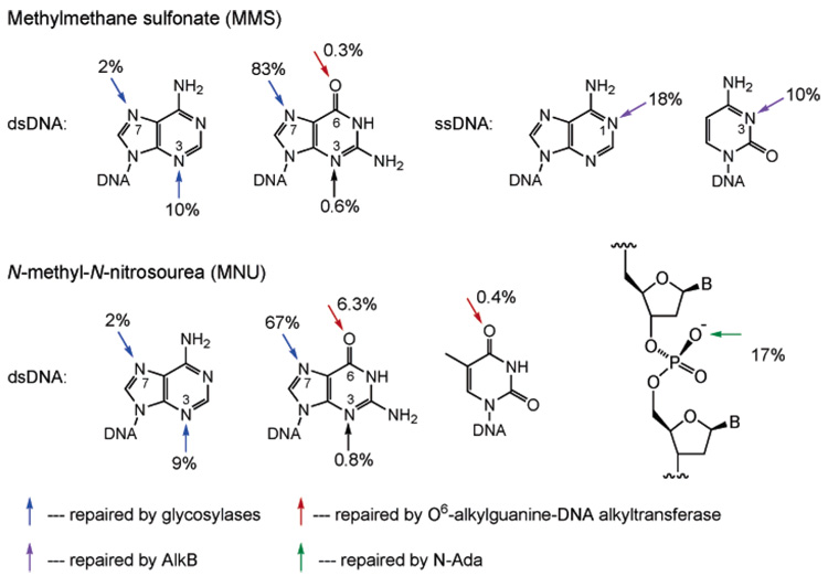
Methylation patterns of the DNA bases and phosphate backbone with MMS and MNU. The blue arrows indicate methylation sites that are repaired by glycosylases; the red arrows are for sites repaired by O6-alkylguanine-DNA alkyltransferases; the purple arrows are for sites repaired by the AlkB proteins; and the green arrow is the site repaired by N-Ada.
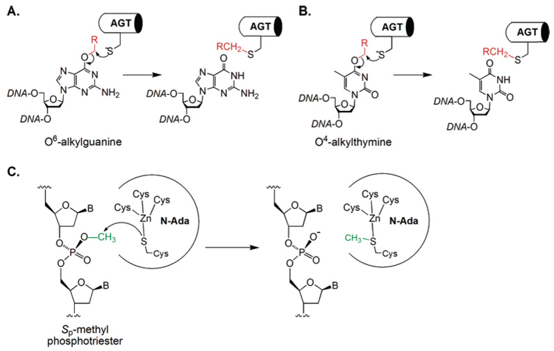
A Cys residue is used by the AGT proteins to remove the alkyl adducts on the O6-position of guanine (A) and the O4-position of thymine (B). A zinc-bound Cys residue is also used by the N-terminal domain of the E. coli Ada protein to displace a methyl group from Sp-methylphosphotriester (C). All these alkyl transfers from DNA lesions to Cys residues are irreversible.
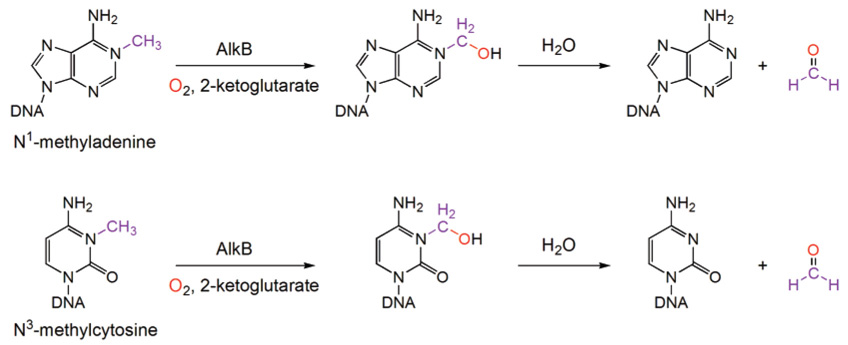
Novel oxidative dealkylation mechanism used by the AlkB proteins to remove methyl or other alkyl groups on the N1-position of adenine and the N3-position of cytosine.
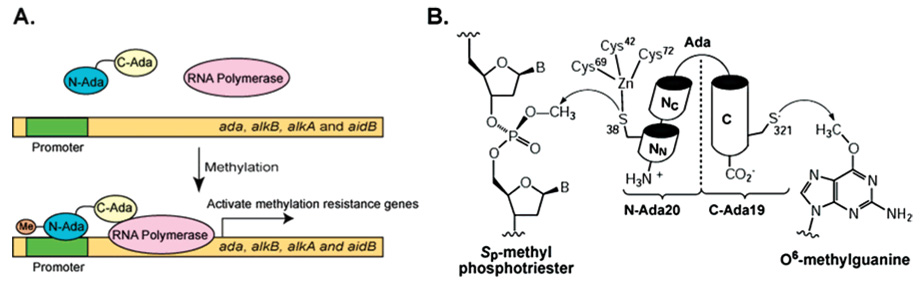
The E. coli Ada protein is a multiple function protein that protects E. coli against methylation challenge. (A) Ada is a transcriptional activator. Methylation of N-Ada increases Ada’s affinity to DNA and converts it into a potent transcriptional activator. N-Methylated Ada binds several promoter sites and recruits RNA polymerase to initiate transcriptional activation of the ada regulon that includes the methylation resistance genes ada, alkB, alkA, and aidB. (B) Ada possesses two repair activities: the N-terminal domain of Ada utilizes a zinc-activated Cys residue to repair Sp-methylphosphotriester; the C-terminal domain contains a nucleophilic Cys residue to remove alkyl groups on the O6-position of guanine and the O4-position of thymine.
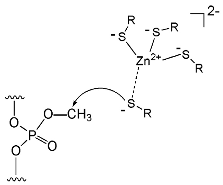
A Cys4Zn cluster accumulates negative charges. A Cys residue, which may transiently dissociate from the zinc(II) center while still kept as the deprotonated form, can attack a methylphos-photriester to transfer the methyl group.
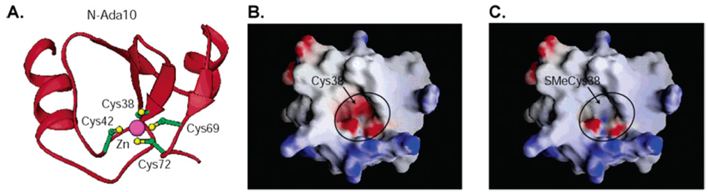
NMR structure of N-Ada10 (PDB accession code 1EYF). (A) The overall fold and the Cys4Zn cluster. (B) An electrostatic surface (GRASP) representation of N-Ada10 in the unmethylated state. Red, negatively charged surfaces; blue, positively charged surfaces; white, neutral. (C) An electrostatic surface (GRASP) representation of N-Ada10 with a methyl group added to the sulfur atom of Cys38 (modeled from the NMR structure). Note the change in charge state of the Cys4Zn pocket that interacts with DNA.
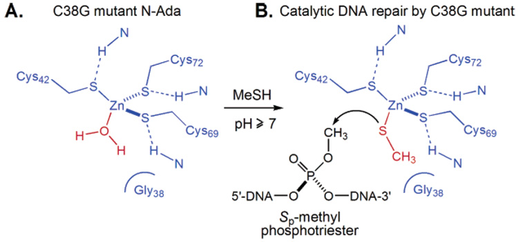
Strategy to convert N-Ada into a catalytic enzyme. (A) The active Cys38 residue is mutated to Gly to open a coordination site on the zinc(II) center. (B) A methanethiol molecule can bind to zinc(II) and perform methyl transfer from Sp-methylphosphotriester.
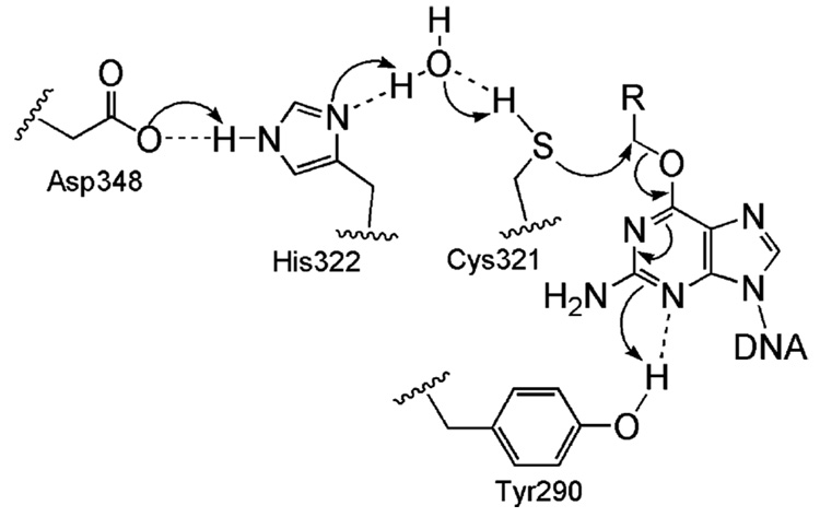
A conserved hydrogen bond network is present in the active site of the AGT proteins to facilitate the repair.
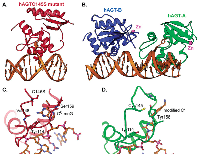
Structures of the hAGT/DNA complexes. (A) Overall structure of an inactive mutant hAGTC145S bound to a dsDNA containing a damaged base O6-meG. The lesioned base is flipped out of the minor groove of the DNA and inserted into the active site of the protein (PDB accession code 1T38). (B) Overall structure of the native hAGT bound to two different regions of DNA: hAGT-A flips a modified cytosine in DNA, and hAGT-B binds two ends of the DNA and recognizes a terminal overhanging T (1YFH). (C) Recognition of the O6-meG in the active site of the hAGTC145S mutant. (D) Binding of the modified cytosine in the active site of the native hAGT-A.
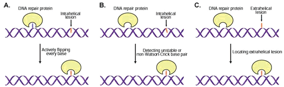
Three potential damage-searching mechanisms for DNA repair proteins that recognize and process damaged bases extrahelically. (A) An active damage-searching mechanism. In this mechanism, the protein flips every base out and checks it in its active site pocket until the lesion is located. (B) A repair protein selectively detects the unstable/non-Watson–Crick base pair that contains the damaged base. (C) A mechanism of simply capturing a transiently extrahelical base lesion by the repair protein.
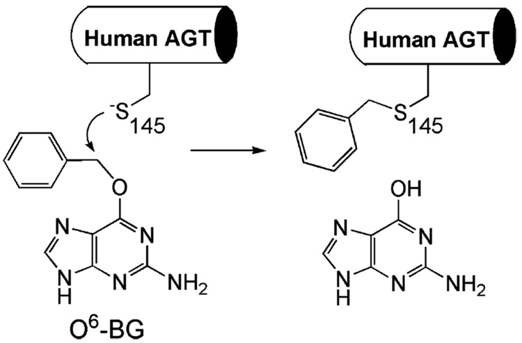
O6-BG irreversibly inactivates hAGT.
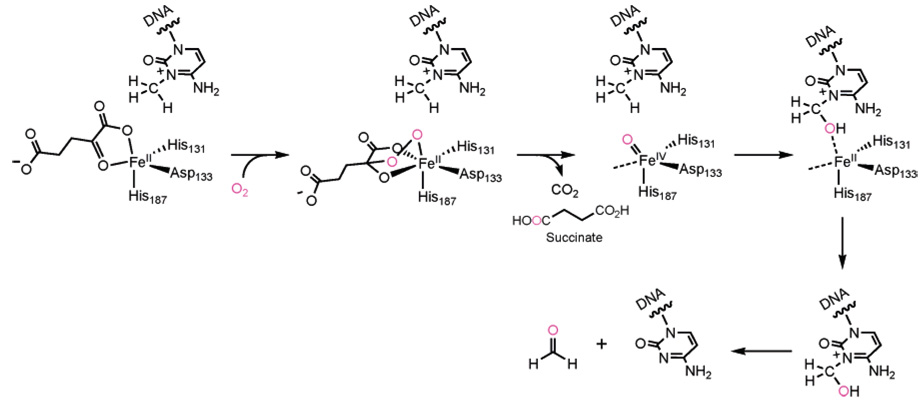
Proposed mechanism for dioxygen activation and dealkylation repair of the AlkB proteins. The O2 reacts with the active site iron(II) and the α-keto carbon of an iron-bound αKG to give a bridged peroxo type intermediate. This intermediate undergoes a concerted decarboxylation and a heterolytic cleavage of the O–O bond to form the key active species, which is speculated to be a high-valent iron(IV)–oxo intermediate. This high-valent species oxidizes the alkyl group (3-meC is shown as an example) to afford an unstable alcohol which decomposes to an aldehyde and the repaired base.
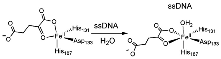
Change of the geometry of the active site iron(II) in E. coli AlkB from 5-coordinate to 6-coordinate with the addition of ssDNA.
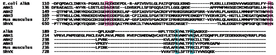
Sequence alignment of several homologues of AlkB, including the first three human homologues (ABH1–3), as well as mouse and viral homologues, generated using ClustalW. The accession numbers of the aligned sequences are NP_311128, NP_006011, NP_001001655, NP_631917, XP-132383, and AAA47787.
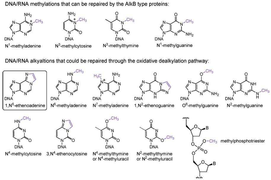
Sites that may also be subject to oxidative dealkylation repair. Exocyclic DNA adducts such as 1,N6-enthenoadenine could be repaired as well.
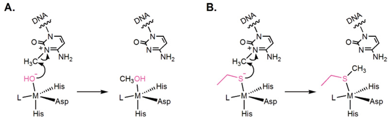
Alternative strategies to repair a 1-meA or 1-meC lesion involving a nucleophile to directly displace the alkyl adduct from the damaged base.
References
-
- Friedberg EC, Walker GC, Siede W. DNA Repair and Mutagenesis. Washington, DC: ASM Press; 1995.
-
- Lindahl T. Nature. Vol. 362. 1993. p. 709. - PubMed
-
- Drabløs F, Feyzi E, Aas PA, Vaagbø CB, Kavli B, Bratlie MS, Peña-Diaz J, Otterlei M, Slupphaug G, Krokan HE. DNA Repair. 2004;3:1389. - PubMed
-
- Vaughan P, Lindahl T, Sedgwick B. Mutat. Res. 1993;293:249. - PubMed
Publication types
MeSH terms
Substances
Grants and funding
LinkOut - more resources
Full Text Sources
