Review
. 2006 Feb;147 Suppl 2(Suppl 2):S132-43.
doi: 10.1038/sj.bjp.0706637.

Purinoceptors as therapeutic targets for lower urinary tract dysfunction

Affiliations
Review

Purinoceptors as therapeutic targets for lower urinary tract dysfunction

Anthony P D W Ford et al. Br J Pharmacol. 2006 Feb.

Abstract

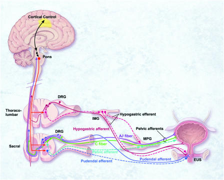
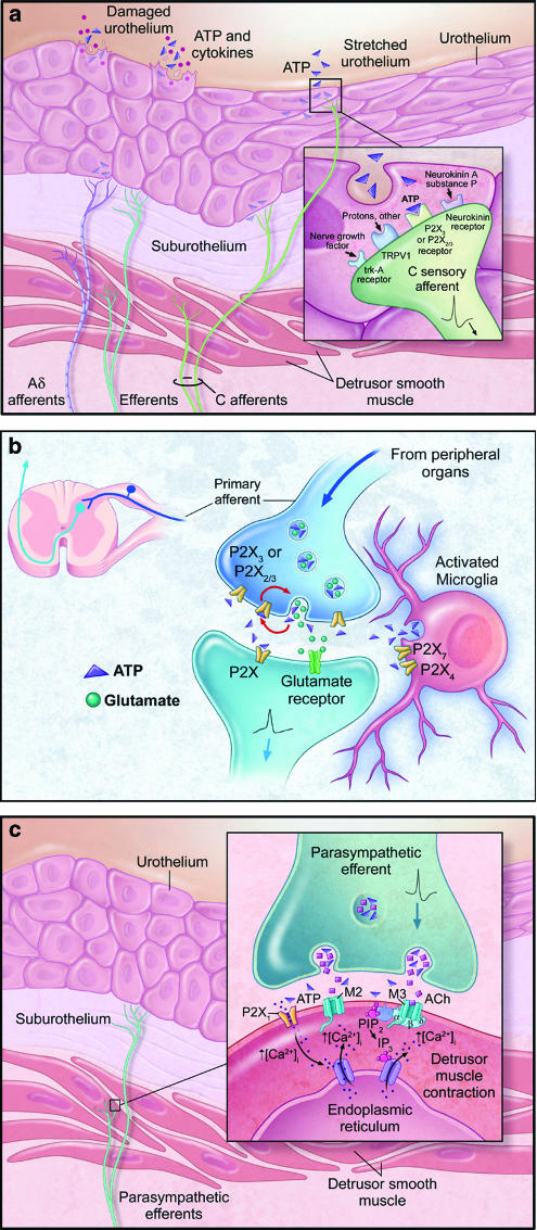
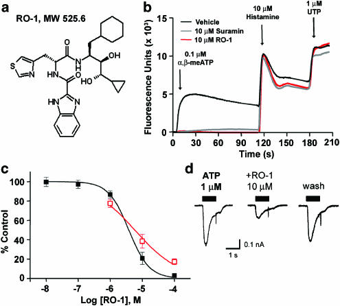
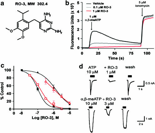
Lower urinary tract symptoms (LUTS) are present in many common urological syndromes. However, their current suboptimal management by muscarinic and alpha(1)-adrenoceptor antagonists leaves a significant opportunity for the discovery and development of superior medicines. As potential targets for such therapeutics, purinoceptors have emerged over the last two decades from investigations that have established a prominent role for ATP in the regulation of urinary bladder function under normal and pathophysiological conditions. In particular, evidence suggests that ATP signaling via P2X(1) receptors participates in the efferent control of detrusor smooth muscle excitability, and that this function may be heightened in disease and aging. ATP also appears to be involved in bladder sensation, via activation of P2X(3) and P2X(2/3) receptors on sensory afferent neurons, both within the bladder itself and possibly at central synapses. Such findings are based on results from classical pharmacological and localization studies in non-human and human tissues, knockout mice, and studies using recently identified pharmacological antagonists--some of which possess attributes that offer the potential for optimization into candidate drug molecules. Based on recent advances in this field, it is clearly possible that the development of selective antagonists for these receptors will occur that could lead to therapies offering better relief of sensory and motor symptoms for patients, while minimizing the systemic side effects that limit current medicines.


Figures

Figure 1
Figure 1
Schematic diagram of the neural circuits controlling continence and micturition. The majority of Aδ- and C-afferents that innervate the urinary bladder and urethra are found in pelvic nerves, which also contain parasympathetic efferents originating from the sacral spinal cord. The remaining bladder afferents are carried by hypogastric nerves, which also contain sympathetic efferents originating from the thoracolumber spinal cord. Sacral somatic afferent and efferent innervation to the external urethral sphincter is via pudendal nerves. Under normal physiological conditions in adults, the micturition reflex is controlled predominantly by Aδ afferents communicating via the spinal cord to supraspinal centers in the pons and cortex. Under pathophysiological conditions or with aging, spinal reflex mechanisms mediated by C-fibre afferents may become dominant.
Figure 2
Figure 2
Schematic diagrams showing the roles of ATP and P2X receptors in the micturition pathway. (a) Mechanical distension or damage to the urothelium causes release of ATP, and this release is augmented in disease states such as interstitial cystitis, benign prostate hyperplasia, or spinal cord injury. ATP acts on P2X3 and P2X2/3 receptors on the peripheral terminals of Aδ- and C-bladder afferents, where it may convey mechanosensory and nociceptive information to the spinal cord. (b) At the central terminals of primary sensory afferents within the dorsal horn of the spinal cord, ATP may be coreleased with glutamate. P2X receptors are expressed on both presynaptic and postsynaptic membranes. Presynaptic P2X3 and P2X2/3 receptors are thought to be important in facilitating glutamate release. In addition, P2X4 and P2X7 receptors present on microglia may mediate inflammatory responses, thus contributing to hyperexcitability at these synapses. (c) Excitation of parasympathetic efferents causes corelease of ATP with acetylcholine from the nerve terminal. These neurotransmitters act on P2X1 and muscarinic (M3) receptors, respectively, present on the postjunctional membrane to cause detrusor smooth muscle contraction.
Figure 3
Figure 3
Structure and in vitro pharmacological properties of RO-1, a selective P2X1 antagonist. (a) Chemical structure of RO-1. (b) Cytosolic calcium flux evoked by 0.1 μM α,β-meATP (first peak), 10 μM histamine (second peak), and 1 μM UTP (third peak) in Fluo-3-loaded CHOK1 cells expressing recombinant human P2X1 receptors. In all, 10 μM suramin and 10 μM RO-1 blocked α,β-meATP-evoked cytosolic calcium flux, but did not inhibit calcium flux evoked by histamine or UTP acting on endogenous histamine and UTP-sensitive, suramin-insensitive P2Y receptors in CHOK1 cells. (c) Concentration-effect curves showing the inhibition of cytosolic calcium flux evoked by 0.1 μM α,β-meATP in Fluo-3-loaded CHOK1 cells expressing recombinant human P2X1 receptors (filled black squares; pIC50=5.5), or currents evoked by 1 μM ATP in patch-clamped, dissociated rat bladder smooth muscle cells (open red squares; pIC50=5.2). (d) Representative patch-clamp recordings from dissociated rat bladder smooth muscle cells showing inhibition of ATP-evoked currents by RO-1. The inhibition of ATP-evoked currents could be reversed following washout of RO-1 from the extracellular bath solution.
Figure 4
Figure 4
Structure and in vitro pharmacological properties of RO-3, a selective P2X3 and P2X2/3 antagonist. (a) Chemical structure of RO-3. (b) Cytosolic calcium flux evoked by 1 μM α,β-MeATP (first peak) and 5 μM ionomycin (second peak) in Fluo-3-loaded CHOK1 cells expressing recombinant rat P2X3 receptors. RO-3 at 0.1 and 1 μM blocked α,β-meATP-evoked cytosolic calcium flux, but did not inhibit calcium flux evoked by ionomycin. (c) Concentration-effect curves showing inhibition of cytosolic calcium flux evoked by 1 μM α,β-meATP in Fluo-3-loaded CHOK1 cells expressing recombinant rat P2X3 receptors (filled black squares; pIC50=7.0) or 5 μM α,β-meATP in Fluo-3-loaded 1321N1 astrocytoma cells expressing recombinant human P2X2/3 receptors (filled black triangles; pIC50=5.9). Also shown are concentration-effect curves for inhibition of currents evoked by 10 μM ATP or α,β-meATP in patch-clamped, dissociated rat thoracolumbar dorsal root ganglion (open red squares; pIC50=6.8) or nodose ganglion neurons (open red triangles; pIC50=5.9), respectively. (d) Representative patch-clamp recordings from dissociated rat thoracolumbar DRG (upper panel) or nodose ganglion (lower panel) neurons showing the inhibition of ATP- or α,β-meATP-evoked currents by RO-3. The inhibition of ATP- or α,β-meATP-evoked currents could be reversed by washout of RO-3 from the extracellular bath solution.

References

    1. ABBRACCHIO M.P., BOEYNAEMS J.M., BARNARD E.A., BOYER J.L., KENNEDY C., MIRAS-PORTUGAL M.T., KING B.F., GACHET C., JACOBSON K.A., WEISMAN G.A., BURNSTOCK G. Characterization of the UDP-glucose receptor (re-named here the P2Y14 receptor) adds diversity to the P2Y receptor family. Trends Pharmacol. Sci. 2003;24:52–55. - PMC - PubMed
    1. ABRAMS P., CARDOZO L., FALL M., GRIFFITHS D., ROSIER P., ULMSTEN U., VAN KERREBROECK P., VICTOR A., WEIN A. The standardisation of terminology of lower urinary tract function: report from the standardisation sub-committee of the International Continence Society. Neurourol. Urodyn. 2002;21:167–178. - PubMed
    1. ANDERSSON K.E. Pharmacology of lower urinary tract smooth muscles and penile erectile tissues. Pharmacol. Rev. 1993;45:253–308. - PubMed
    1. ANDERSSON K.E., WEIN A. Pharmacology of the lower urinary tract: basis for current and future treatments of urinary incontinence. Pharmacol. Rev. 2004;56:581–631. - PubMed
    1. BARCLAY J., PATEL S., DORN G., WOTHERSPOON G., MOFFATT S., EUNSON L., ABDEL'AL S., NATT F., HALL J., WINTER J., BEVAN S., WISHART W., FOX A., GANJU P. Functional downregulation of P2X3 receptor subunit in rat sensory neurons reveals a significant role in chronic neuropathic and inflammatory pain. J. Neurosci. 2002;22:8139–8147. - PMC - PubMed

MeSH terms