

Molecular mechanisms of detrusor and corporal myocyte contraction: identifying targets for pharmacotherapy of bladder and erectile dysfunction
- PMID: 16465183
- PMCID: PMC1751499
- DOI: 10.1038/sj.bjp.0706627
Molecular mechanisms of detrusor and corporal myocyte contraction: identifying targets for pharmacotherapy of bladder and erectile dysfunction
Abstract
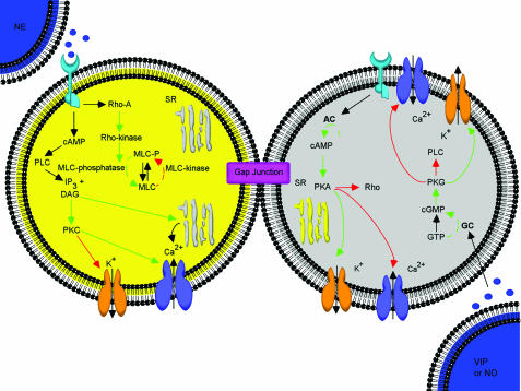
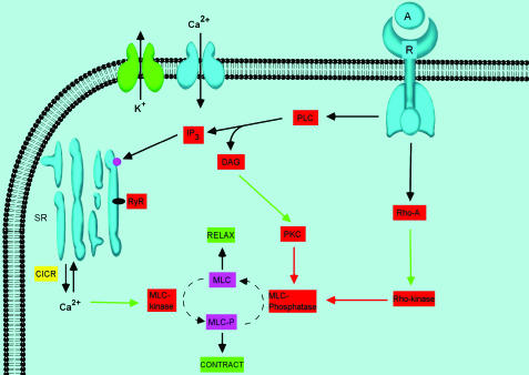
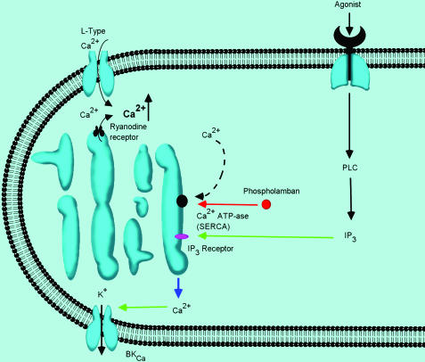
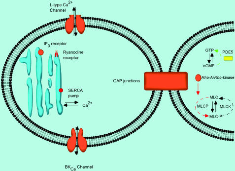
The Post-Genomic age presents many new challenges and opportunities for the improved understanding, diagnosis and treatment of human disease. The long-term goal is to identify molecular correlates of disease processes, and use this information to develop novel and more effective therapeutics. A major hurdle in this regard is ensuring that the molecular targets of interest are indeed relevant to the physiology and/or pathophysiology of the processes being studied, and, moreover, to determine if they are specific to the tissue/organ being investigated. As a first step in this direction, we have reviewed the literature pertaining to bladder and erectile physiology/pharmacology and dysfunction and attempted to summarize some of the critical molecular mechanisms regulating detrusor and corporal myocyte tone. Because of the vast amount of published data, we have limited the scope of this review to consideration of the calcium-mobilizing and calcium-sensitizing pathways in these cells. Despite obvious differences in phenotypic characteristics of the detrusor and corporal myocyte, there are some common molecular changes that may contribute to, for example, the increased myocyte contractility characteristic of bladder and erectile dysfunction (i.e. increased Rho kinase activity and decreased K(+) channel function). Of course, there are also some important distinctions in the pathways that modulate contractility in these two cell types (i.e. the contribution of ryanodine-sensitive calcium stores and the nitric oxide/cGMP pathways). This report highlights some of these similarities and distinctions in the hope that it will encourage scientific discourse and research activity in this area, eventually leading to an improved quality of life for those millions of individuals that are afflicted with bladder and erectile dysfunction.
Figures
References
-
- ANDERSSON K.E. Neurophysiology/pharmacology of erection. Int. J. Impot. Res. 2001;13 (Suppl 3):S8–S17. - PubMed
-
- ANDERSSON K.E.2003Erectile physiological and pathophysiological pathways involved in erectile dysfunction J. Urol. 170S6–S13.discussion S13–S14. - PubMed
-
- ANDERSSON K.E. Detrusor contraction – focus on muscarinic receptors. Scand. J. Urol. Nephrol. Suppl. 2004;215:54–57. - PubMed
-
- ANDERSSON K.E., ARNER A. Urinary bladder contraction and relaxation: physiology and pathophysiology. Physiol. Rev. 2004;84:935–986. - PubMed
-
- ARCHER S.L. Potassium channels and erectile dysfunction. Vasc. Pharmacol. 2002;38:61–71. - PubMed
Publication types
MeSH terms
Grants and funding
LinkOut - more resources
Full Text Sources
Other Literature Sources
Medical
Research Materials
