. 2006 Mar;78(3):510-9.
doi: 10.1086/500850. Epub 2006 Jan 19.

Loss-of-function mutations in the keratin 5 gene lead to Dowling-Degos disease

Affiliations

Loss-of-function mutations in the keratin 5 gene lead to Dowling-Degos disease

Regina C Betz et al. Am J Hum Genet. 2006 Mar.

Abstract

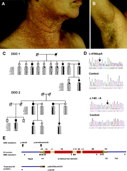
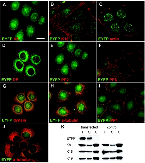
Dowling-Degos disease (DDD) is an autosomal dominant genodermatosis characterized by progressive and disfiguring reticulate hyperpigmentation of the flexures. We performed a genomewide linkage analysis of two German families and mapped DDD to chromosome 12q, with a total LOD score of 4.42 ( theta =0.0) for marker D12S368. This region includes the keratin gene cluster, which we screened for mutations. We identified loss-of-function mutations in the keratin 5 gene (KRT5) in all affected family members and in six unrelated patients with DDD. These represent the first identified mutations that lead to haploinsufficiency in a keratin gene. The identification of loss-of-function mutations, along with the results from additional functional studies, suggest a crucial role for keratins in the organization of cell adhesion, melanosome uptake, organelle transport, and nuclear anchorage.


Figures

Figure  1
Figure 1
Clinical appearance and underlying genetic defect of DDD. A and B, Reticulate hyperpigmentation and hyperkeratotic dark-brown papules on the neck and the axilla of individual II:13 (pedigree DDD 1). C, Two pedigrees segregating for DDD. Marker haplotypes on chromosome 12q13.11-12q15 that are linked to DDD are indicated by black bars. D, Sequence analysis showing the c.418dupA and c.14C→A mutations in patients with DDD. E, Domain structure and position of mutations in K5. Head and tail domains are marked in blue. Conserved helical end domains are marked in green, and nonhelical linker domains L1, L12, and L2 are marked with yellow bars. 1A, 1B, 2A, and 2B indicate rod subdomains. The thin red line in coil 2B indicates the “stutter.” H1 and H2 are conserved α-helical subdomains flanking the rod. The head domain extends from aa 1 to 132, H1 from 133 to 158, 1A from 159 to 203, 1B from 216 to 316, 2A from 334 to 352, 2B from 360 to 481, H2 from 481 to 501, and the tail domain from 502 to 590. The positions of the mutations p.Ser5X (c.14C→A) and p.Ile140AsnfsX39 (c.418dupA) are indicated by black arrowheads. Mutations causing EBS are marked by black lines. Detailed information about the localization of EBS mutations is available at the Human Intermediate Filament Mutation Database. The residual K5 polypeptides are generated by the p.Ser5X and p.Ile140AsnfsX39 mutations. The truncated K5 protein caused by p.Ile140AsnfsX39 encompasses positions 1–139 of K5 and 39 aa of an incorrect reading frame (gray bar).
Figure  2
Figure 2
Light microscopy and Fontana-Masson staining of sections from DDD and control skin. A, Hematoxylin and eosin staining of sections from DDD demonstrates papillary epidermal downgrowth, in contrast to control skin (B). C, Fontana-Masson staining shows scattered distribution of melanosomes in patients with DDD. D, Regular, nuclear caplike structure of melanosomes in control skin. Scale bar represents 50 μm.
Figure  3
Figure 3
Electron microscopy of skin from an individual with DDD and from a control. A, Semithin sections from a patient with DDD with filiform epithelial downgrowth (arrows), in contrast to control skin (B). Note irregular DDD cell shape and size in (A). C–G, Ultrathin sections from a patient with DDD with scattered distribution of melanosomes (E) and altered perinuclear organization of keratins in suprabasal but not in basal cells. C and D, Perinuclear rim of filament-free, smooth cytoplasm is demarcated by arrowheads; SC = spinous cell. K5 haploinsufficiency allows formation of normal keratin filaments and their interaction with hemidesmosomes (F) and desmosomes (G). Scale bars represent 20 μm (A and B), 25 μm (C), 500 nm (D and E), and 200 nm (F and G).
Figure  4
Figure 4
Immunofluorescence analysis of skin sections. Skin sections from a patient with DDD (A and C) and from an unaffected individual (B and D) stained with antibodies against K5 and K14 (A and B) and K10 (C and D) revealed no differences in the distribution and organization of keratins. Note the presence of terminally differentiated cells in the papillary downgrowth. The dashed lines in panels C and D indicate the basal lamina. Scale bar represents 50 μm.
Figure  5
Figure 5
Immunofluorescence and biochemical analysis of transfected MCF-7 and HaCaT cells stably expressing an EYFP-p.Ile140fs fusion protein. A and B, MCF-7 cells showing random distribution of the K5 head domain throughout transfectants, including nuclei. Staining for K18, representative of endogenous keratins, revealed no colocalization of EYFP-p.Ile140fs with endogenous keratins (B). Stably transfected MCF-7 cells are stained with antibodies against actin (C), desmoplakin (DP) (D), plakophilin 2 (PP2) (E), and plakophilin 3 (PP3) (F). Stably transfected HaCaT keratinocytes are stained with antibodies against plakophilin 1 (PP1) (I) and α-tubulin (J). In both cell lines, no colocalization or altered distribution between EYFP-p.Ile140fs and any of these proteins was detected. In MCF-7 cells, a partial colocalization of EYFP-p.Ile140fs (yellow overlay) with dynein intermediate chain (G), but not with α-tubulin (H), was detected. Fractionation and immunoblotting of MCF-7 cells stably transfected with EYFP-p.Ile140fs and mock-transfected controls (K). Blots containing total (T), soluble (S), and Triton/high-salt–insoluble cytoskeletal proteins (C) were incubated with antibodies against EYFP, K8, K18, and K19. EYFP-p.Ile140fs was detected exclusively in the soluble fraction harvested from transfected MCF-7 cells, without affecting the solubility of endogenous keratins. Scale bar represents 20 μm.

Similar articles

Cited by

References

Web Resources

    1. GenBank, http://www.ncbi.nlm.nih.gov/Genbank/ (for KRT5 [accession numbers NM_000424 and NP_000415])
    1. Human Intermediate Filament Mutation Database, http://www.interfil.org/ - PubMed
    1. Online Mendelian Inheritance in Man (OMIM), http://www.ncbi.nlm.gov/Omim/ (for DDD) - PubMed

References

    1. Biltz H, Kiessling M (1988) Dowling-Degos disease: an autosomal dominant genetic dermatosis. Z Hautkr 63:642–644 - PubMed
    1. Braun-Falco M, Ring J (2003) Enhanced cytoplasmic expression of desmocollin 3 in epidermal rete ridges of Dowling-Degos syndrome. Br J Dermatol 149:1293–129610.1111/j.1365-2133.2003.05645.x - DOI - PubMed
    1. Braun-Falco M, Volgger W, Borelli S, Ring J, Disch R (2001) Galli-Galli disease: an unrecognized entity or an acantholytic variant of Dowling-Degos disease? J Am Acad Dermatol 45:760–76310.1067/mjd.2001.116340 - DOI - PubMed
    1. Byers HR, Maheshwary S, Amodeo DM, Dykstra SG (2003) Role of cytoplasmic dynein in perinuclear aggregation of phagocytosed melanosomes and supranuclear melanin cap formation in human keratinocytes. J Invest Dermatol 121:813–82010.1046/j.1523-1747.2003.12481.x - DOI - PubMed
    1. Chang L, Goldman RD (2004) Intermediate filaments mediate cytoskeletal crosstalk. Nat Rev Mol Cell Biol 5:601–61310.1038/nrm1438 - DOI - PubMed

Publication types

Associated data