. 2006 Feb;13(2):227-34.
doi: 10.1128/CVI.13.2.227-234.2006.

Human interleukin-15 improves engraftment of human T cells in NOD-SCID mice

Affiliations

Human interleukin-15 improves engraftment of human T cells in NOD-SCID mice

Anyuan Sun et al. Clin Vaccine Immunol. 2006 Feb.

Abstract

Human nonobese diabetic-severe combined immune deficiency (NOD-SCID) mouse chimeras have been widely used as an in vivo model to assess human immune function. However, only a small fraction of transferred human T lymphocytes can be detected in human peripheral blood lymphocyte (huPBL)-NOD-SCID chimeras. To improve the reconstitution of human T lymphocytes in NOD-SCID mice, the use of recombinant human interleukin-15 (rhIL-15) as a stimulator of human lymphocytes was explored. Administration of rhIL-15 after transplantation of huPBLs into NOD-SCID mice increased reconstitution of human T lymphocytes in a dose-dependent manner, with an optimal dosage of 1 microg/mouse. The number of human T lymphocytes (HLA-ABC+ CD3+) in the lymphoid organs or tissue of rhIL-15-treated huPBL-NOD-SCID mice increased 11- to 80-fold, and phytohemagglutinin-induced T-lymphocyte proliferation and cytokine production were significantly enhanced. Additionally, although mature human cells have not been thought to enter the murine thymus, human T lymphocytes were detected in the huPBL-NOD-SCID thymus after rhIL-15 treatment. Thus, rhIL-15 can be used to optimize long-term peripheral T-cell engraftment in these human-mouse chimeras and may also be useful in clinical treatment of T-cell deficiencies.


Figures

FIG. 1.
FIG. 1.
Protocol for examination of engraftment of huPBLs into NOD-SCID mice. huPBLs were separated by Ficoll density gradient centrifugation and washed twice with HBSS. After the cells were counted and adjusted to 1 × 10⁸/ml, 5 × 10⁷/0.5 ml of the purified huPBLs were transferred i.p. into recipient NOD-SCID mice that had been pretreated with irradiation. After transfer, the huPBL-NOD-SCID mice were injected i.p. either with the indicated amount of rhIL-15 or with HBSS as a control every other day for a total of 10 injections starting on day 1.
FIG. 2.
FIG. 2.
rhIL-15 promoted the transplantation of human T cells into lymphoid organs of NOD-SCID mice in a dose-dependent manner. Recipient mice that had been pretreated with irradiation received transplants of freshly prepared huPBLs. To determine the optimal dosage of rhIL-15, the huPBL-NOD-SCID mice were then divided into five groups with five mice in each group. Four dosages of rhIL-15 in HBSS were tested in this study: 0.25, 0.5, 1, and 2 μg of rhIL-15 in 0.2 ml of HBSS per mouse, and an equal volume of HBSS was used in control groups. Mice were injected i.p. either with different dosages of rhIL-15 or with HBSS every other day for a total of 10 injections. Four weeks after the huPBL transplantation, mononuclear cells from the thymus and spleen were isolated and stained with anti-HLA-ABC, anti-CD3, and anti-CD19 and were then analyzed by flow cytometry. Data are representative of three independent experiments, with similar results obtained in each experiment. P < 0.001 for comparison between rhIL-15 groups with different dosages and the HBSS group.
FIG. 3.
FIG. 3.
rhIL-15 accelerated human T-lymphocyte engraftment into lymphoid organs or tissues of NOD-SCID mice. One microgram of rhIL-15 in 0.2 ml of HBSS or an equal volume of HBSS as a control was injected i.p. into each huPBL-NOD-SCID mouse once. The injection scheme of rhIL-15 or HBSS is diagramed in Fig. 1. One week after the last rhIL-15 injection (on day 28), the organs or tissues, including the peritoneal cavity, thymus, spleen, liver, lymph nodes, and peripheral blood, were collected, and the mononuclear cells from the organs or tissues were isolated as described in Materials and Methods, stained with PE-labeled anti-HLA-ABC and Cy-labeled anti-CD3, and then analyzed by flow cytometry. (A) HLA-ABC+ and CD3+ percentages of the lymphoid organs or tissues from grafted mice administered HBBS and rhIL-15. Data shown are representative of three independent experiments, with similar results obtained in each experiment. P < 0.01 for comparison of the rhIL-15 group with the HBSS group. (B) HLA-ABC+ CD3+ cell populations were calculated from total-lymphoid-organ cell numbers and 1 ml peripheral blood of the mice using the proportions (percentages) of these cells. Results are expressed as means ± standard deviations for three independent assays. P < 0.001 for comparison of the rhIL-15 group with the HBSS group.
FIG. 4.
FIG. 4.
rhIL-15 promoted the trafficking of human T cells to lymphoid organs of NOD-SCID mice. To prove the promoting role of IL-15 in T-cell trafficking to lymphoid organs and tissues, we collected the mononuclear cells from the peritoneal cavities, spleens, and thymuses of rhIL-15-treated huPBL-NOD-SCID mice on days 7, 14, 21, and 28 after huPBL transfer, stained them with PE-labeled anti-HLA-ABC and Cy-labeled anti-CD3, and then analyzed them by flow cytometry. Data are representative of three independent experiments, with similar results obtained in each experiment.
FIG. 5.
FIG. 5.
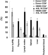
Flow cytometry analysis of human T-lymphocyte subsets from lymphoid organs or tissues of huPBL-NOD-SCID mice. To test human T-lymphocyte subsets from lymphoid organs or tissues of huPBL-NOD-SCID mice, on day 28, the lymphoid organs or tissues of huPBL-NOD-SCID mice and donor huPBLs were collected, stained with Cy-labeled anti-CD3, FITC-labeled anti-CD4, and PE-labeled anti-CD8 antibodies, and then analyzed by flow cytometry. The analysis was performed on gated lymphocytes with FSC/SSC characteristics. Results are expressed as means ± standard deviations for three independent assays. P < 0.001 for comparison of the rhIL-15 group with the HBSS group.
FIG. 6.
FIG. 6.
Proliferation of the cells of lymph nodes from rhIL-15-treated huPBL-NOD-SCID chimeras stimulated with PHA in vitro. On day 28 after huPBL transfer, the cells of lymph nodes of rhIL-15-treated mice and donor huPBLs were harvested. All cells (1 × 10⁵ human T cells/100 μl/well) were cultured in 96-well flat-bottom plates with or without 10 μg/ml PHA. Three days later, proliferation activity was assayed by pulsing with 1 μCi (3.7 × 10⁴ Bq) of [3H]thymidine (6.7 Ci/mmol) for 8 h; the radioactivity for each sample was counted by a liquid scintillation counter. Data are presented as the means ± standard deviations of triplicate samples. There was no obvious difference between donor T cells and rhIL-15-treated chimera human T cells in response to PHA. P < 0.001 for comparison of the lymphocytes of the rhIL-15-treated chimera with and without PHA.
FIG. 7.
FIG. 7.
Cytokine production by the cells of lymph nodes from huPBL-NOD-SCID mice treated with rhIL-15. The cells of lymph nodes from rhIL-15-treated mice and donor huPBLs were used in this experiment. The cells were cultured in 96-well flat-bottom plates with or without 10 μg/ml PHA in vitro. At 18, 36, or 72 h, as indicated, cell culture supernatants were collected for determination of IFN-γ (A) or IL-2 (B) by quantitation ELISA. Data are presented as the means ± standard deviations of triplicate samples. There was no obvious difference in cytokine production between donor T cells and rhIL-15-treated chimera human T cells. P < 0.001 for comparison of the lymphocytes of the rhIL-15-treated chimera at different time points with and without PHA.

References

    1. Abedi, M. R., B. Christensson, K. B. Islam, L. Hammarstrom, and C. I. E. Smith. 1992. Immunoglobulin production in severe combined immunodeficient (SCID) mice reconstituted with human peripheral blood mononuclear cells. Eur. J. Immunol. 22:823-828. - PubMed
    1. Albert, S. E., C. Mckerlie, A. Pester, B.-J. Edgell, J. Carlyle, M. Petric, and J. W. Chamberlain. 1997. Time-dependent induction of protective antiinfluenza immune responses in human peripheral blood lymphocyte/SCID mice. J. Immunol. 159:1393-1403. - PubMed
    1. Baird, A. M., R. M. Gerstein, and L. J. Berg. 1999. The role of cytokine receptor signaling in lymphocyte development. Curr. Opin. Immunol. 11:157-166. - PubMed
    1. Barry, T. S., D. M. Jones, C. B. Richter, and B. F. Haynes. 1991. Successful engraftment of human postnatal thymus in severe combined immune deficient (SCID) mice: differential engraftment of thymic components with irradiation versus anti-asialo GM-1 immunosuppressive regimens. J. Exp. Med. 173:167-180. - PMC - PubMed
    1. Bernasconi, N. L., E. Traggiai, and A. Lanzavecchia. 2002. Maintenance of serological memory by polyclonal activation of human memory B cells. Science 298:2199-2202. - PubMed

Publication types