. 2006 Mar 22;25(6):1375-84.
doi: 10.1038/sj.emboj.7600969. Epub 2006 Feb 9.

Structure of glutamate carboxypeptidase II, a drug target in neuronal damage and prostate cancer

Affiliations

Structure of glutamate carboxypeptidase II, a drug target in neuronal damage and prostate cancer

Jeroen R Mesters et al. EMBO J. .

Abstract

Membrane-bound glutamate carboxypeptidase II (GCPII) is a zinc metalloenzyme that catalyzes the hydrolysis of the neurotransmitter N-acetyl-L-aspartyl-L-glutamate (NAAG) to N-acetyl-L-aspartate and L-glutamate (which is itself a neurotransmitter). Potent and selective GCPII inhibitors have been shown to decrease brain glutamate and provide neuroprotection in preclinical models of stroke, amyotrophic lateral sclerosis, and neuropathic pain. Here, we report crystal structures of the extracellular part of GCPII in complex with both potent and weak inhibitors and with glutamate, the product of the enzyme's hydrolysis reaction, at 2.0, 2.4, and 2.2 A resolution, respectively. GCPII folds into three domains: protease-like, apical, and C-terminal. All three participate in substrate binding, with two of them directly involved in C-terminal glutamate recognition. One of the carbohydrate moieties of the enzyme is essential for homodimer formation of GCPII. The three-dimensional structures presented here reveal an induced-fit substrate-binding mode of this key enzyme and provide essential information for the design of GCPII inhibitors useful in the treatment of neuronal diseases and prostate cancer.


Figures
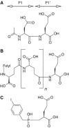
Figure 1
Figure 1
Chemical formulae for (A) N-acetyl-L-aspartyl-L-glutamate (NAAG), (B) folyl-poly-γ-glutamate, (C) (S)-2-(4-iodobenzylphosphonomethyl)-pentanedioic acid (GPI-18431). The P1 and P1′ moieties are indicated for (A).
Figure 2
Figure 2
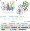
Structure of GCPII. (A, B) Three-dimensional structure of the dimer. One subunit is shown in gray, while the other is colored according to organization into domains. Domain I, light blue; domain II, yellow; domain III, brown. The dinuclear zinc cluster at the active site is indicated by dark green spheres, the Ca2+ ion near the monomer–monomer interface by a red sphere, and the Cl ion by a yellow sphere. The GPI-18431 inhibitor is shown as small beige balls. The ‘glutarate sensor' (the β15/β16 hairpin) is shown in light green. The seven carbohydrate side chains located in the electron density maps are indicated. The position of the structure relative to the membrane is shown in (A). (B) Provides a view into the ‘cup' of the dimeric enzyme (see text). The entrance to the catalytic site is indicated (‘E'). (C) Primary and secondary structure of GCPII, colored according to domain organization. Domain I, light blue; domain II, yellow; domain III, brown.
Figure 3
Figure 3
Surface representation of the ∼20 Å deep funnel leading to the catalytic site. Blue, side-chain nitrogens of Arg and Lys residues; red, side-chain oxygens of Asp and Glu; green, side-chain carbons of Tyr and Phe residues. Yellow, Zn2+ ions; inhibitors shown as stick models. (A) Complex with GPI-18431; (B) complex with phosphate. Note the difference in the shape of the pocket because of withdrawal of the ‘glutarate sensor' (Y700) in the phosphate complex.
Figure 4
Figure 4
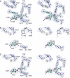
2FoFc electron density maps (stereo) contoured at 1.2σ, for the GCPII complex with GPI-18431 (A), phosphate (B), and L-glutamate (C). Zinc ions are shown in dark green, chloride in yellow. Ligands are shown using green sticks and atom-color spheres. Note the different conformation of the ‘glutarate sensor' (Lys699 and Tyr700) in (B), which is caused by the absence of a glutarate moiety in the phosphate complex.
Figure 5
Figure 5
Scheme of the catalytic site, indicating the NAAG substrate as bound to the enzyme. The binding of the NAA portion of the substrate to the S1 pocket (left) is modeled, whereas the interactions of the glutamate residue to the S1′ pocket (right) are taken from the crystal structure of the glutamate complex (cf. Figure 4C). Residues Lys699 and Tyr700 of the ‘glutarate sensor' are boxed. The first step in the catalytic reaction could be activation of the central nucleophile, HO–H, through the general base, Glu424. This is followed by the nucleophilic attack of the generated hydroxyl ion onto the peptidic bond of the substrate. After cleavage, Glu424 shuttles the proton to the amino group of the leaving product. Arrows run from nucleophile to electrophile.

References

    1. Anilkumar G, Rajasekaran SA, Wang S, Hankinson O, Bander NH, Rajasekaran AK (2003) Prostate-specific membrane antigen association with filamin A modulates its internalization and NAALADase activity. Cancer Res 63: 2645–2648 - PubMed
    1. Barinka C, Rinnova M, Sacha P, Rojas C, Majer P, Slusher BS, Konvalinka J (2002) Substrate specificity, inhibition and enzymological analysis of recombinant human glutamate carboxypeptidase II. J Neurochem 80: 477–487 - PubMed
    1. Barinka C, Sacha P, Sklenar J, Man P, Bezouska K, Slusher BS, Konvalinka J (2004) Identification of the N-glycosylation sites on glutamate carboxypeptidase II necessary for proteolytic activity. Protein Sci 13: 1627–1635 - PMC - PubMed
    1. Bennett MJ, Lebron JA, Bjorkman PJ (2000) Crystal structure of the hereditary haemochromatosis protein HFE complexed with transferrin receptor. Nature 403: 46–53 - PubMed
    1. Benveniste H, Drejer J, Schousboe A, Diemer NH (1984) Elevation of the extracellular concentrations of glutamate and aspartate in rat hippocampus during transient cerebral ischemia monitored by intracerebral microdialysis. J Neurochem 43: 1369–1374 - PubMed

Publication types

MeSH terms