. 2005:3A:325-340.

Glioma cell integrin expression and their interactions with integrin antagonists: Research Article

Glioma cell integrin expression and their interactions with integrin antagonists: Research Article

Ralph-Heiko Mattern et al. Cancer Ther. 2005.

Abstract

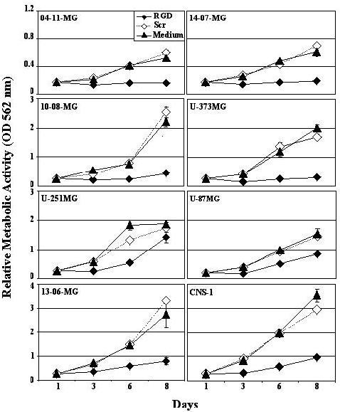
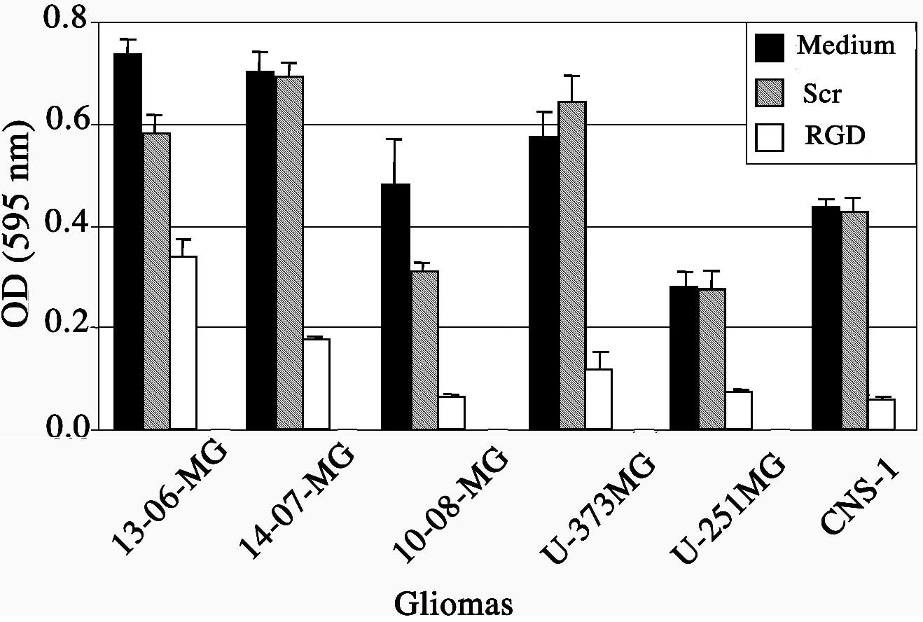
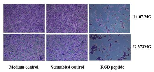
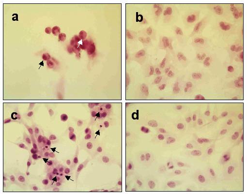
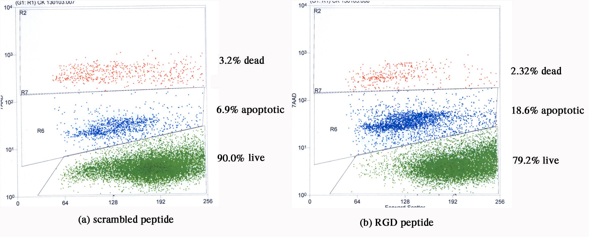
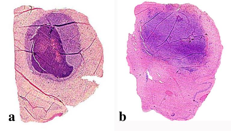
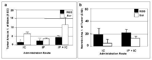
A panel of human glioma cell explants was screened for integrin expression by flow cytometry using α(ν)β-specific antibodies. A lower percentage of the glioma cells were positive for the α(ν)β3 (mean % positive = 20.8%) integrin, whereas higher percentages were positive for the ανβ5 (mean % positive = 72.7%), VLA5α (mean % positive = 87%) and VLAβ1 (mean % positive = 41.7%) integrins. A series of RGD peptides was designed, synthesized and tested for binding to integrin receptors. Based on the results of the binding to the isolated integrin receptors and the expression of integrins on glioma cell lines, a peptide that binds potently to the α(ν)β3, α(ν)β5 and α(5)β(1) was selected for further investigations with regards to its effect on glioma cells. The peptide, Ac-c[(Pen)-Tyr(Me)-Ala-Arg-Gly-Asp-Asn-Tic-Cys]NH(2) (RGD peptide), exhibited high potential for use in clinical intracranial administration since it had good stability in rat brain cell homogenates placed into artificial cerebrospinal fluid. Using an HPLC method for quantification of peptides in rat brain cell homogenates, we could demonstrate the half-life of the RGD peptide approximated 20 hr. Relative to a scrambled peptide control (non-RGD sequence, same amino acids), the experimental RGD peptide significantly decreased glioma cell proliferation of the entire panel of rat and human glioma cells tested. Adhesion of recently passaged glioma cells to glioma-derived extracellular matrix protein-coated plates was inhibited significantly by the RGD peptide. The peptide also reversed attachment of plated glioma cells. The RGD peptide caused some, but not substantial, glioma cell injury, as evidenced by a quantitative in vitro nuclear DNA morphologic assay and by a flow cytometric assay employing 7-amino actinomycin D (7AAD). We histologically monitored for toxicity caused by various doses of the RGD peptide infused repeatedly into normal cannulated rat brain. At safe doses, the experimental RGD peptide-treated brains did not show significant differences from those infused with scrambled peptide or buffer-treated controls. In tumor-bearing brains, slightly smaller tumor areas were measured with a higher necrotic-to-tumor index in the RGD peptide treated relative to the scrambled peptide-treated controls. This was obtained with intracranial peptide administrations or combined intracranial and intraperitoneal injections. From this in vitro work, we conclude that the anti-glioma effects of the RGD peptide tested resulted from lowered glioma proliferation and adhesion/mobility, rather than from significant glioma cell injury in the timeframe analyzed. Although other mechanisms not discerned from our limited histopathological observations may be operational, from our in vivo work, we conclude that repeated administration of RGD peptide into brain is safe but that better delivery of the peptides to infiltrating tumor cells is necessary.


Figures

Figure 1
Figure 1
RGD peptide effects on glioma cell proliferation. A panel of glioma cells were assayed when incubated with fresh medium, or that containing scrambled non-RGD peptide (Scr) or RGD peptide. The relative metabolic activity was determined by an in vitro colorimetric method. The mean of the absorbances obtained at 562 nm from supernates harvested from triplicate wells ± SE is shown at 1, 3, 6 and 8 days.
Figure 2
Figure 2
RGD peptide effects on adhesion of glioma cells to autologous glioma cell extracts of extracellular matrix proteins. The mean absorbance values ± SE obtained at 595 nm were from triplicate wells containing crystal violet stained cells. Cells were incubated for 4 hr with fresh medium (■) or that containing scrambled peptide () or RGD peptide (□).
Figure 3
Figure 3
Crystal violet stained 14-07-MG and U-373MG glioma cells adhered to ECM protein-coated wells after 4 hr incubation in fresh medium, in medium containing scrambled peptide, or RGD peptide.
Figure 4
Figure 4
Quantitative morphologic assay showing H&E stained brain tumor cell monolayers that were or were not exposed to RGD peptide (a) human 10-08-MG glioma cells after 18 hr coincubation with RGD peptide demonstrate fewer attached cells and rounded shapes. Some of the cells exhibit typical apoptotic morphological changes such as those with condensed nuclei (black arrow) and fragmented DNA (white arrow) (b) 10-08-MG glioma cells not exposed to RGD peptide exhibited fewer apoptotic cells. The cells were larger with oval nuclei and abundant cytoplasm (c) rat 9L gliosarcoma cells after 4 hr coincubation with RGD peptide. Apoptotic cells with condensed nuclei (black arrows) are visible in the monolayer (d) 9L gliosarcoma cells in monolayer and not exposed to peptide. The healthy cells are well attached to the surface and mitotic figures are readily apparent.
Figure 5
Figure 5
Scattergrams from a 7AAD flow cytometric assay, which give live, apoptotic and dead (necrotic/late apoptotic) cell percentages after a short 3 hr treatment of rat 9L gliosarcoma cells with (a) scrambled non-RGD peptide and (b) RGD peptide. An approximate 2-fold increase in injured 9L gliosarcoma cells is seen when incubated with RGD peptide compared to the scrambled counterpart. Forward scatter (abscissa) is plotted versus 7AAD intensity (ordinate).
Figure 6
Figure 6
Photomicrographs of RGD or scrambled peptide repeatedly introduced intracranially into cannulated normal rat brain (a-c) or 9L tumor-bearing rat brain (d-i). At 24 hr following the last infusion: (a) Rat brains administered RGD peptide at high doses show signs of stress including congested vessels (black arrows) and capillaries (white arrow). (b) At higher power, brains given RGD peptide at higher doses also showed infiltration of mononuclear cells and polymorphonuclear cells (black arrows), indicative of an acute inflammatory reaction. At 7 or 14 days following the last infusion: (c) Small focal sterile granulomas often formed in cannulated rat brain, whether treated with saline or peptide, as visualized by a necrotic center (Nc) surrounded by a fibrotic wall (Fb). Normal brain (NB) was immediately adjacent to the granuloma. (d) At low power, a representative sized area of necrosis characteristic of scrambled peptide treated brains is shown next to (e) a representative larger sized area of necrosis characteristic of RGD peptide treated brains. The (f) saline-treated tumor has viable cells showing a number of mitotic figures (black arrows) and (g) tumor cells at the periphery of the solid tumor mass are infiltrating into perivascular spaces within normal brain. The (h) RGD peptide treated brain sections show many cells with pyknotic nuclei interspersed within the solid tumor mass adjacent to the instillation site (i) often the palisading areas of growth better show the pyknotic cells that are assumed undergoing apoptosis. Healthy tumor cells are also shown growing in perivascular spaces of the RGD peptide treated brains, however, similar to that shown in g. Magnifications are: d,e = 40X, a,c = 100X, h,i = 200X, f,g = 400X, b = 600X
Figure 7
Figure 7
H&E stained sections macroscopically showing tumor filling the majority of the upper part of the hemispheres. Panel (a) shows a tumor containing a substantial degree of necrosis derived from an animal that was treated with IC, IP RGD peptide. Panel (b) shows a tumor not displaying necrosis that was derived from an animal that was treated with IC, IP scrambled peptide. Magnifications are 4X
Figure 8
Figure 8
Tumor areas as percentages of total brain areas at the instillation site and necrotic areas as percentages of the total tumor areas, evaluated in groups of RGD and scrambled (Scr) peptide treated rat brains. The peptides were infused 6 times over a two week period. Sacrifice was at 48 hr following the last infusion. (a) The CNS-1 tumor areas are given as percentages of total brain area when administration of RGD or scrambled peptides was by intracranial (IC) or intraperitoneal (IP) routes, or both (IP + IC). (b) Necrotic areas as percentages of the tumor areas at the instillation site when administered by IC or IP + IC routes. The tumors given peptides by the IP route were not necrotic (data not shown).* By unpaired t test with Welch correction, comparing RGD vs Scr given IC the p value was significant at 0.0263. Also, comparing RGD given IP vs IP+IC was not considered quite significant with p=0.0547.
None
Carol A. Kruse

References

    1. Bartus RT, Elliott PJ, Dean RL, Hayward NJ, Nagle TL, Huff MR, Snodgrass PA, Blunt DG. Controlled modulation of BBB permeability using the bradykinin agonist, RMP-7. Exp Neurol. 1996;142:14–28. - PubMed
    1. Berens ME, Giese A. What's malignant about astrocytomas? Studies of brain tumor proliferation and migration. Barrows Neurol Inst Quarterly. 1996;12:15–22.
    1. Buckley CD, Pilling D, Henriquez NV, Parsonage G, Threlfall K, Scheel-Toellner D, Simmons DL, Akbar AN, Lord JM, Salmon M. RGD peptides induce apoptosis by direct caspase-3 activation. Nature. 1999;397:534–9. - PubMed
    1. CBTRUS . Central Brain Tumor Registry of the United States. 2002. CBTRUS: Statistical Report: Primary Brain Tumors in the United States, 1995-1999.
    1. Chatterjee S, Matsumura A, Schradermeier J, Gillespie GY. Human malignant glioma therapy using anti-α(v)β3 integrin agents. J Neurooncol. 2000;46:135–44. - PubMed

LinkOut - more resources