Case Reports
. 2006 Oct;55(10):1185-97.
doi: 10.1007/s00262-005-0118-2. Epub 2006 Feb 9.

Tumor progression despite massive influx of activated CD8(+) T cells in a patient with malignant melanoma ascites

Affiliations
Case Reports

Tumor progression despite massive influx of activated CD8(+) T cells in a patient with malignant melanoma ascites

Helena Harlin et al. Cancer Immunol Immunother. 2006 Oct.

Abstract

Although melanoma tumors usually express antigens that can be recognized by T cells, immune-mediated tumor rejection is rare. In many cases this is despite the presence of high frequencies of circulating tumor antigen-specific T cells, suggesting that tumor resistance downstream from T cell priming represents a critical barrier. Analyzing T cells directly from the melanoma tumor microenvironment, as well as the nature of the microenvironment itself, is central for understanding the key downstream mechanisms of tumor escape. In the current report we have studied tumor-associated lymphocytes from a patient with metastatic melanoma and large volume malignant ascites. The ascites fluid showed abundant tumor cells that expressed common melanoma antigens and retained expression of class I MHC and antigen processing machinery. The ascites fluid contained the chemokines CCL10, CCL15, and CCL18 which was associated with a large influx of activated T cells, including CD8(+) T cells recognizing HLA-A2 tetramer complexes with peptides from Melan-A and NA17-A. However, several functional defects of these tumor antigen-specific T cells were seen, including poor production of IFN-gamma in response to peptide-pulsed APC or autologous tumor cells, and lack of expression of perforin. Although these defects were T cell intrinsic, we also observed abundant CD4(+)CD25(+)FoxP3(+) T cells, as well as transcripts for FoxP3, IL-10, PD-L1/B7-H1, and indoleamine-2,3-dioxygenase (IDO). Our observations suggest that, despite recruitment of large numbers of activated CD8(+) T cells into the tumor microenvironment, T cell hyporesponsiveness and additional negative regulatory mechanisms can limit the effector phase of the anti-tumor immune response.


Figures

Fig. 1
Fig. 1
Real-time RT-PCR of total ascites cells and of the cell line established from ascites. cDNA was analyzed for expression of melanoma tumor antigens gp100, MAGE-3, Melan-A, and NA17-A, and for β-actin by real-time RT-PCR. a Real-time RT-PCR from total ascites cells. b Real-time RT-PCR from the melanoma cell line
Fig. 2
Fig. 2
Lymphocyte subset analysis and T cell activation status. a The percent of lymphocytes among total ascites and peripheral blood cells was determined by flow cytometry (top panel). Among these lymphocytes, the percent of cells staining positive for CD19 (B cells) and CD3 (T cells and NK T cells) were analyzed (middle panel), as well as the percent of cells that were CD56+ CD3 (NK cells), CD56+ CD3+ (NK T cells), and CD56 CD3+ (T cells) (bottom panel). b The percent of activated T cells was determined by staining for MHC class II (HLA-DR) expression among either the CD8+ (top panel) or CD4+ (bottom panel) CD3+ cells
Fig. 3
Fig. 3
Flow cytometric analysis of intracellular cytokine production. a Ascites cells were either left unstimulated (resting) or stimulated with PMA + ionomycin (activated), surface stained, and permeabilized for intracellular staining. They were then incubated with antibodies against either IL-2 (upper panels), IL-4 (middle panels), or IFN-γ (lower panels) prior to analysis by flow cytometry. The percent CD8+ and CD8 cells producing each cytokine out of the total pool of CD8+ or CD8 cells were determined and are indicated in the dot plots. b Peripheral blood cells were treated and analyzed in the same manner, and in parallel with, the ascites cells in panel a
Fig. 4
Fig. 4
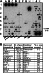
Cytokine/chemokine array of ascites fluid. Ascites fluid was analyzed for the presence of 79 different cytokines and chemokines using a membrane spotted with antibodies directed against the cytokines and chemokines. After blocking the membrane, it was incubated with undiluted ascites fluid, then washed and incubated with a cocktail of biotin-conjugated secondary antibodies. After further washing and incubation with HRP-conjugated streptavidin, the presence of bound cytokine was detected using ECL and the membrane exposed to film. a Image of the membrane, showing the cytokine and chemokine spots. b Densitometry analysis, showing the relative pixel intensity compared to the positive control spots (1A–D, 8J–K), adjusted for the negative control spots (1E–F, 8H)
Fig. 5
Fig. 5
Analysis of chemokine receptor expression of the total ascites cells and on the cell line. RNA extracted from the cells was analyzed for expression of chemokine receptors by RT-PCR, as compared to control cDNA expressing all of the genes analyzed. GAPDH was used as a control for cDNA quality, and RNA prepared using no RT (RT−) used to control for genomic DNA contamination. a RNA from total ascites or cell line analyzed for expression of CXCR1-4 and the chemokine SDF-1α. b RNA from total ascites cells or the melanoma cell line analyzed for expression of CCR1-5
Fig. 6
Fig. 6
Tetramer analysis of antigen-specific CD8+ T cells. a Mononuclear cells from ascites fluid were stained with the indicated tetramers and anti-CD8 antibody. The percent tetramer+ CD8+ cells among all CD8+ cells were determined and is shown. b Peripheral blood cells were stained and analyzed in the same manner as, and in parallel with, the ascites cells in panel a
Fig. 7
Fig. 7
Phenotypic analysis of CD8+ T cells in the ascites. Total CD8+ T cells were analyzed for expression of CD45RA, CD62L, CD27, and CD28. EBV, Melan-A, and NA17-A-specific CD8+ T cells, as determined by tetramer staining, were also gated on and analyzed for the expression of these surface markers. The percent cells found in each quadrant or gate (shown in the dot plots) for total CD8+ T cells as compared to the tetramer-specific CD8+ T cells are shown in table format
Fig. 8
Fig. 8
Antigen presentation in ascites tumor cells. a HLA-A2 expression by tumor cells. Tumor cell line cells were stained with FITC anti-HLA-A2 or isotype control and analyzed by flow cytometry. b RT-PCR of antigen presentation pathway components. cDNA (RT+) or mock cDNA made in the absence of RT (RT−) from normal donor PBMC, the tumor cell line, or TAP deficient T2 cells was analyzed for expression of TAP1, TAP2, LMP2, LMP7, and LMP10. 1 normal donor RT+, 2 normal donor RT−; 3 tumor cell line RT+, 4 tumor cell line RT−; 5 T2 cells RT+, 6 T2 cells RT−. c Purified CD8+ T cells from a normal donor were cultured together with tumor cell line cells that were either not loaded with peptide or pulsed with EBV peptide. The culture was performed in normal medium or in the presence of 50% ascites fluid. The number of IFN-γ spots/105 CD8+ T cells plated is shown. d T cell lines specific for Melan-A and gp100 peptides were stimulated with the patient’s tumor cell line and ELISpot analysis was performed. Stimulation with K562-A2 cells is shown as a control
Fig. 9
Fig. 9
Functional analysis of antigen-specific CD8+ T cells. a Purified CD8+ T cells from ascites fluid were cultured together with T2 cells that were either not loaded with peptide or pulsed with EBV peptide, or with the melanoma tumor cell line cells. b CD8+ T cells from the ascites were cultured in the presence of T2 cells loaded with the indicated peptides or PMA + ionomycin for 24 h. The number of IFN-γ spots was analyzed for each well, and is shown as the number of spots/105 total cells. c Perforin expression of antigen-specific T cells. Ascites cells were surface stained with antibodies against CD8 and with tetramers and then stained for intracellular perforin. Lymphocytes were gated on by size and granularity and are shown, with the percent of cells stained with EBV, Melan-A, and NA17-A tetramers, on the left. The perforin staining is shown on the right, with percent cells in each quadrant indicated in each dot plot
Fig. 10
Fig. 10
Negative regulatory factors present in the ascites. a Real-time RT-PCR of total ascites cells. RNA extracted from the cells was analyzed for expression of Arginase I, FoxP3, IDO, PD-L1, and for β-actin by real-time RT-PCR. b Results for each of the transcripts shown in a were quantitated for relative expression compared to β-actin. c Ascites cells were surface stained with antibodies against CD3, CD4, and CD25 and permeabilized to stain with anti-FoxP3. Analysis was gated on CD3+ CD4+ cells that were either CD25high or CD25low, and analyzed for the percent FoxP3+ cells

Similar articles

Cited by

References

    1. Rosenberg SA. Progress in human tumour immunology and immunotherapy. Nature. 2001;411:380. doi: 10.1038/35077246. - DOI - PubMed
    1. Marincola FM, Wang E, Herlyn M, Seliger B, Ferrone S. Tumors as elusive targets of T-cell-based active immunotherapy. Trends Immunol. 2003;24:335. doi: 10.1016/S1471-4906(03)00116-9. - DOI - PubMed
    1. Delp K, Momburg F, Hilmes C, Huber C, Seliger B. Functional deficiencies of components of the MHC class I antigen pathway in human tumors of epithelial origin. Bone Marrow Transplant. 2000;25(Suppl 2):S88. doi: 10.1038/sj.bmt.1702363. - DOI - PubMed
    1. Peterson AC, Harlin H, Gajewski TF. Immunization with Melan-A peptide-pulsed peripheral blood mononuclear cells plus recombinant human interleukin-12 induces clinical activity and T-cell responses in advanced melanoma. J Clin Oncol. 2003;21:2342. doi: 10.1200/JCO.2003.12.144. - DOI - PubMed
    1. Plebanski M, Allsopp CE, Aidoo M, Reyburn H, Hill AV. Induction of peptide-specific primary cytotoxic T lymphocyte responses from human peripheral blood. Eur J Immunol. 1995;25:1783. doi: 10.1002/eji.1830250645. - DOI - PubMed

Publication types

MeSH terms