. 2006;8(2):R37.
doi: 10.1186/ar1891. Epub 2006 Jan 16.

CD95-induced osteoarthritic chondrocyte apoptosis and necrosis: dependency on p38 mitogen-activated protein kinase

Affiliations

CD95-induced osteoarthritic chondrocyte apoptosis and necrosis: dependency on p38 mitogen-activated protein kinase

Lei Wei et al. Arthritis Res Ther. 2006.

Abstract

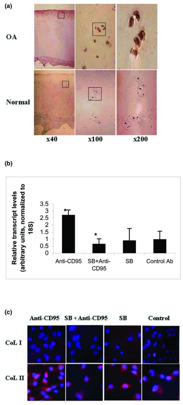
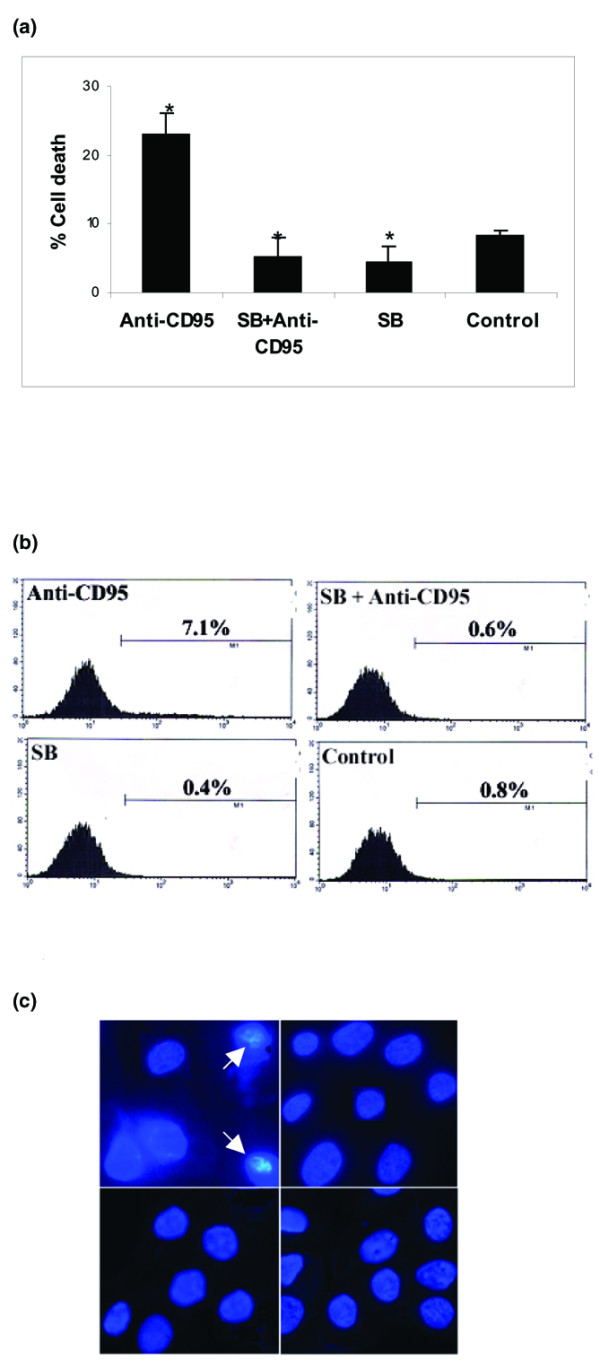
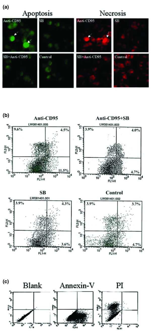
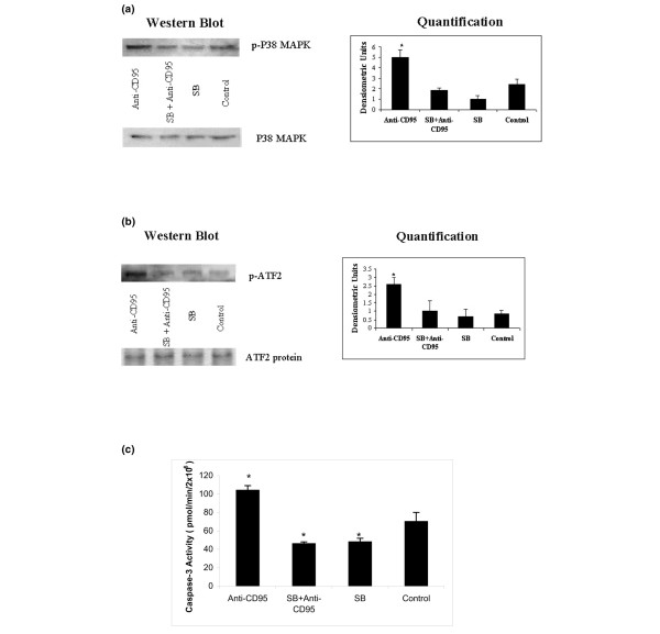
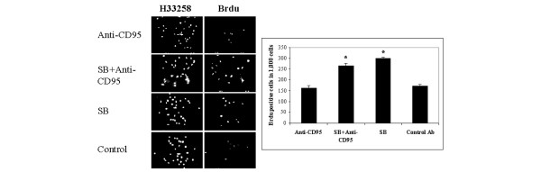
One of the hallmarks of osteoarthritic cartilage is the loss of chondrocyte cellularity due to cell death. However, considerable controversy has recently arisen surrounding the extent of apoptotic cell death involved in development of osteoarthritis (OA). To shed light on this issue, we characterized cell death in primary OA chondrocytes mediated by the CD95 (Fas) pathway. Treatment of chondrocytes with anti-CD95 not only increased the rate of cell death but also increased the production of CD95 ligand by chondrocytes. This reveals a novel autocrine regulatory loop whereby activated chondrocytes may amplify CD95 signals by inducing synthesis of CD95 ligand. Multiple morphologic detection analyses indicated that apoptosis accounted for only a portion of chondrocyte death, whereas the other chondrocytes died by necrosis. Both chondrocyte apoptosis and necrosis depended on the activity of p38 mitogen-activated protein kinase (MAPK) within chondrocytes. Treatment of chondrocytes with the p38 MAPK inhibitor SB203580 abolished anti-CD95 induced cell death by inhibiting the activities of activating transcription factor-2 and caspase-3. In addition, inhibition of p38 MAPK activity in chondrocytes stimulated chondrocyte proliferation, as indicated by 5-bromo-2-deoxyuridine (BrdU) index. Thus, p38 MAPK is a potential therapeutic target, inhibition of which may maintain the cellularity of articular chondrocytes by inhibiting cell death and its amplification signal and by increasing cell proliferation.


Figures

Figure 1
Figure 1
Expression of CD95 and CD95L by OA chondrocytes. (a) Micrographs of immunohistochemcal analysis of CD95 expression in normal (bottom panels) and OA cartilage (top panels). Frozen sections from normal and OA articular cartilage were incubated with a monoclonal antibody CH 11 against CD95. Different (original) magnifications are indicated. Results are representative of two normal and four OA cartilage samples. (b) mRNA levels of CD95L in primary OA chondrocytes by real-time RT-PCR analysis. Total RNA was isolated from chondrocytes from OA cartilage following treatment as indicated below. *P < 0.05 versus treatment with Control Ab (a mouse isotype control antibody IgM). Each bar represents the mean ± standard deviation of three experiments. OA chondrocytes (donor age 63 years) stained with annexin V and PI. Results are representative of three OA cartilage samples. (c) Micrographs of immunocytochemical analysis of collagens type II and type I in OA chondrocytes following treatment as indicated below. Chondrocytes were reacted with rhodomaine mAb against type II or type I collagen, respectively. Chondrocyte nuclei were indicated by Hoechest blue dye staining. Anti-CD95: treatment with a mAb anti-Fas CH 11 (100 ng/ml) in serum-free medium for 17 hours. SB: treatment with SB203580 (10 μmol/l) for 17 hours. SB+Anti-CD95: treatment with SB203580 for 2 hours followed by anti-Fas treatment for 17 hours. Control: cells were treated with DMSO only. Results are representative of 3 OA cartilage samples. CD95L, CD95 ligand; DMSO, dimethyl sulfoxide; mAb, monoclonal antibody; OA, osteoarthritis; PI, propidium iodide.
Figure 2
Figure 2
p38 MAPK activity regulates cell death induced by anti-CD95. (a) Total cell death rate for OA chondrocytes quantified by trypan blue exclusion assay after treatment of cells as indicated below. *P < 0.05 versus control. The graph shows the average of three independent experiments. (b) Apoptosis rate of OA chondrocytes quantified by flow cytometry. Chondrocytes were fluorescently labeled by TUNEL assay after treatment of cells as indicated below. The data shown are representative of three OA cartilage samples. Anti-CD95: treatment with a mAb anti-Fas CH 11 (100 ng/ml) in serum-free medium for 17 hours. SB: treatment with SB203580 (10 μmol/l) for 17 hours. SB+Anti-CD95: treatment with SB203580 for 2 hours followed by anti-Fas treatment for 17 hours. Control: cells were treated with DMSO only. (c) Chondrocytes were labeled with Hoechst after treatment of cells as indicated above. Nuclear condensation (indicated by an arrow) was detected in the cells treated with Anti-CD95. DMSO, dimethyl sulfoxide; mAb, monoclonal antibody; MAPK, mitogen-activated protein kinase; OA, osteoarthritis; PI, propidium iodide; TUNEL, terminal dUTP nick-end labeling.
Figure 3
Figure 3
Chondrocyte death consists of apoptosis and necrosis. (a) Morphologic analysis of chondrocyte apoptosis and necrosis. The nuclei of apoptotic cells were labeled by TUNEL with green fluorescence whereas the nuclei of necrotic cells were labeled by PI with red fluorescence (donor age 63 years). All cell nuclei were labeled with blue Hoechst dye. Results are representative of three OA cartilage samples. (b) The rates of cell apoptosis and necrosis quantified by flow cytometry. Apoptotic cells were labeled by annexin V whereas necrotic cells were labeled by PI at the same time. Total events are 10,000. The x axis represents FL1-H (log) with green fluorchrome for annexin V labeling, and the Y axis represents FL2-H (log) with red fluorchrome for PI labeling. The percentage of cells that are single or double positive for annexin V and PI is indicated in each grid. The upper left grid represents the number of PI single positive cells. The lower right grid represents the number of annexin V single positive cells. The upper right grid represents the number of cells positive for both PI and annexin V. The number of cells that are single positive for PI is 9.6% for anti-CD95, and 3.9% for control, SB, and Anti-CD95+SB. Anti-CD95: treatment with a mAb anti-Fas CH 11 (100 ng/ml) in serum-free medium for 17 hours. SB: treatment with SB203580 (10 μmol/l) for 17 hours. SB+Anti-CD95: treatment with SB203580 for 2 hours followed by anti-Fas treatment for 17 hours. Control: cells were treated with DMSO only. Results are representative of five OA cartilage samples. (c) FACS analysis of OA chondrocytes labeled by annexin V and PI distinguishes apoptosis from necrosis. Chondrocytes were labeled into four groups by Annexin-V-FLUOS Staining Kit: x axis is FL1-H (log), and fluorchrome is green for annexin V (panel b [lower right] and panel c [lower right]); and y axis is FL2-H (log), and fluorchrome is red for PI (panel b [upper left] and panel c [upper left]). Living cells are shown to the lower left and double staining to the upper right of panels b and c. DMSO, dimethyl sulfoxide; FACS, fluorescence-activated cell sorter; mAb, monoclonal antibody; OA, osteoarthritis; PI, propidium iodide; TUNEL, terminal dUTP nick-end labeling.
Figure 4
Figure 4
Anti-CD95 induced chondrocyte death is p38 MAPK dependent. (a) Western blot analysis of the levels of phosohorylated-p38 MAPK and p38 MAPK protein (left panel) and the quantification of the western blot by densitometric analysis (right panel). *P < 0.05 versus control. Each bar represents mean ± standard deviation of three experiments. (b) The levels of phosphorylated ATF-2 and ATF-2 protein were determined by western blot on the left. Quantification of the western blot by densitometric analysis is shown on the right. *P < 0.05 versus control. Each bar represents mean ± standard deviation of three experiments. (c) Caspase-3 activities were determined using a caspase-3 cellular activity assay kit. *P < 0.05 versus control. each bar represents mean ± standard deviation of three experiments. Anti-CD95: treatment with a mAb anti-Fas CH 11 (100 ng/ml) in serum-free medium for 17 hours. SB: treatment with SB203580 (10 μmol/l) for 17 hours. SB+Anti-CD95: treatment with SB203580 for 2 hours followed by anti-Fas treatment for 17 hours. Control: cells were treated with DMSO only. Results are representative of three OA cartilage samples. ATF, activating transcription factor; DMSO, dimethyl sulfoxide; mAb, monoclonal antibody; MAPK, mitogen-activated protein kinase; OA, osteoarthritis.
Figure 5
Figure 5
Cell proliferation regulated by p38 MAPK activity. (a) The number of BrdU positive cells in 1,000. (b) Cell proliferation was evaluated by BrdU staining whereas all cell nuclei were stained by Hoechst 33258 dye. *P < 0.05 versus control. Each bar represents mean ± standard deviation of three experiments. Anti-CD95: treatment with a mAb anti-Fas CH 11 (100 ng/ml) in serum-free medium for 17 hours. SB: treatment with SB203580 (10 μmol/l) for 17 hours. SB+Anti-CD95: treatment with SB203580 for 2 hours followed by anti-Fas treatment for 17 hours. Control Ab: cells were treated with a mouse isotype control antibody IgM. Results are representative of three OA cartilage samples. BrdU, 5-bromo-2-deoxyuridine; mAb, monoclonal antibody; MAPK, mitogen-activated protein kinase; OA, osteoarthritis.

References

    1. Bendele AM, Hulman JF. Spontaneous cartilage degeneration in guinea pigs. Arthritis Rheum. 1988;31:561–565. - PubMed
    1. Vignon E, Arlot M, Patricot LM, Vignon G. The cell density of human femoral head cartilage. Clin Orthop Relat Res. 1976;121:303–308. - PubMed
    1. Wei L, Brismar BH, Hultenby K, Hjerpe A, Svensson O. Distribution of chondroitin 4-sulfate epitopes (2/B/6) in various zones and compartments of articular cartilage in guinea pig osteoarthrosis. Acta Orthop Scand. 2003;74:16–21. doi: 10.1080/00016470310013590. - DOI - PubMed
    1. Blanco FJ, Guitian R, Vazquez-Martul E, de Toro FJ, Galdo F. Osteoarthritis chondrocytes die by apoptosis. A possible pathway for osteoarthritis pathology. Arthritis Rheum. 1998;41:284–289. doi: 10.1002/1529-0131(199802)41:2<284::AID-ART12>3.0.CO;2-T. - DOI - PubMed
    1. Hashimoto S, Ochs RL, Komiya S, Lotz M. Linkage of chondrocyte apoptosis and cartilage degradation in human osteoarthritis. Arthritis Rheum. 1998;41:1632–1638. doi: 10.1002/1529-0131(199809)41:9<1632::AID-ART14>3.0.CO;2-A. - DOI - PubMed

Publication types

MeSH terms