

Association between microscopic brain damage as indicated by magnetization transfer imaging and anticardiolipin antibodies in neuropsychiatric lupus
- PMID: 16469116
- PMCID: PMC1526597
- DOI: 10.1186/ar1892
Association between microscopic brain damage as indicated by magnetization transfer imaging and anticardiolipin antibodies in neuropsychiatric lupus
Abstract
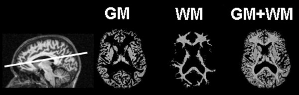
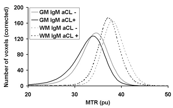
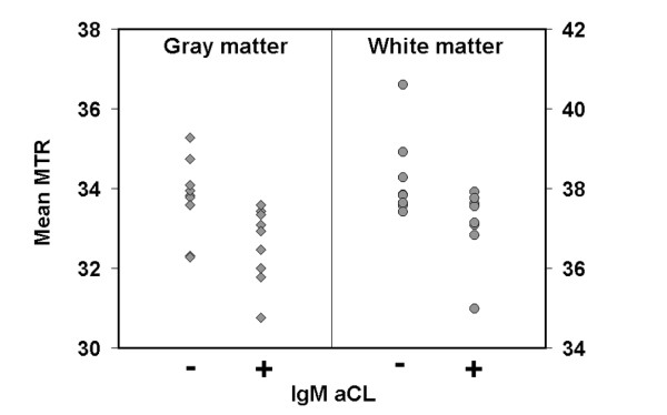
The pathogenetic role of anticardiolipin antibodies (aCLs) in patients with neuropsychiatric systemic lupus erythematosus (NPSLE) without cerebral infarcts remains elusive. Magnetization transfer imaging (MTI) has proved to be a sensitive tool for detecting diffuse microscopic brain damage in NPSLE patients. In this study we examined the correlation between grey and white matter magnetization transfer ratio (MTR) parameters and the presence of IgM and IgG aCLs and lupus anticoagulant in 18 patients with systemic lupus erythematosus and a history of NPSLE but without cerebral infarcts on conventional magnetic resonance imaging. Lower grey matter mean MTR (P < 0.05), white matter mean MTR (P < 0.05), white matter peak location (P < 0.05) and grey matter peak location (trend toward statistical significance) were observed in IgM aCL-positive patients than in IgM aCL-negative patients. No significant differences were found in MTR histogram parameters with respect to IgG aCL and lupus anticoagulant status, nor with respect to anti-dsDNA or anti-ENA (extractable nuclear antigen) status. This is the first report of an association between the presence of aCLs and cerebral damage in grey and white matter in NPSLE. Our findings suggest that aCLs are associated with diffuse brain involvement in NPSLE patients.
Figures
References
-
- Tofts PS, Steens SC, van Buchem MA. Quantitative MRI of the Brain: Measuring Changes Caused by Disease. Chichester, UK: Wiley; 2003. MT: magnetization transfer; pp. 257–298.
MeSH terms
Substances
LinkOut - more resources
Full Text Sources
Medical
