. 2006 Feb 10;124(3):631-44.
doi: 10.1016/j.cell.2005.11.043.

p120-catenin mediates inflammatory responses in the skin

Affiliations

p120-catenin mediates inflammatory responses in the skin

Mirna Perez-Moreno et al. Cell. .

Abstract

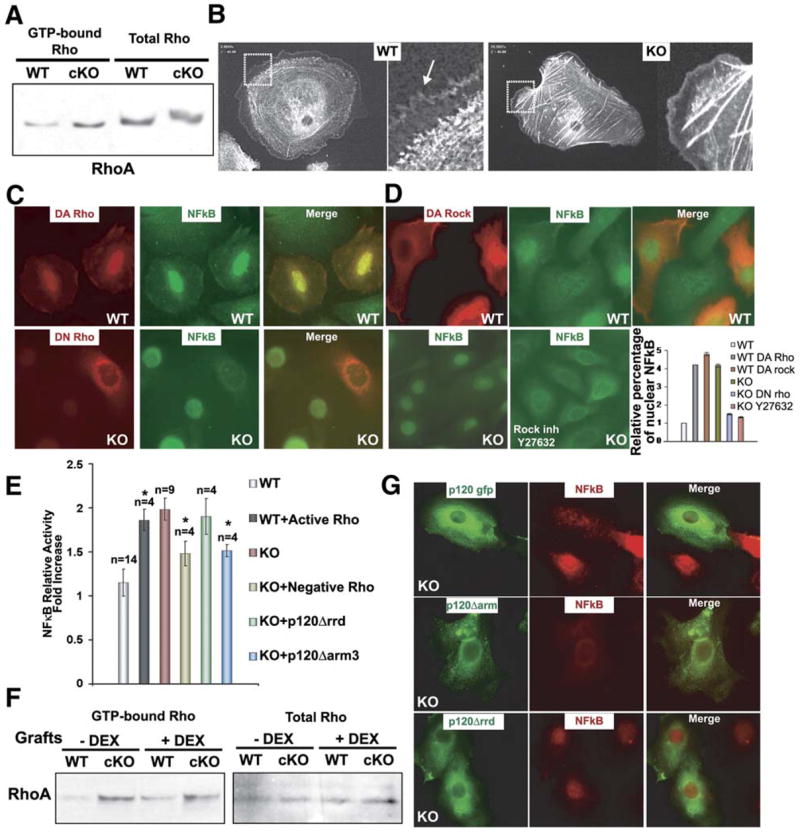
Although p120-catenin regulates adherens junction (AJ) stability in cultured cells, genetic studies in lower eukaryotes have not revealed a role for this protein in vivo. Using conditional targeting in mice, we show that p120 null neonatal epidermis exhibits reduced intercellular AJ components but no overt disruption in barrier function or intercellular adhesion. As the mice age, however, they display epidermal hyperplasia and chronic inflammation, typified by hair degeneration and loss of body fat. Using skin engraftments and anti-inflammatory drugs, we show that these features are not attributable to reductions in junctional cadherins and catenins, but rather NFkB activation. Both in vivo and in vitro, p120 null epidermal cells activate nuclear NFkB, triggering a cascade of proinflammatory NFkB targets. Although the underlying mechanism is likely complex, we show that p120 affects NFkB activation and immune homeostasis in part through regulation of Rho GTPases. These findings provide important new insights into p120 function.


Figures

Figure 1
Figure 1. Phenotypic and Biochemical Alterations in Newborn Mice Conditionally Null for p120 in Skin Epidermis
(A) PCR analysis to confirm genotype. All PCR fragments were of the expected sizes. (B) P0 mice. (C and D) Outside-in (β-galactosidase, blue) and inside-out (biotin, red) dye penetration assays on E18.5mice to test epidermal integrity. Sections are counterstained with anti-occludin to verify tight junction integrity and barrier exclusion. sc, stratum corneum. (E) Ultrastructural analysis of basal epidermal layer. Insets are magnified views of boxed areas. Intercellularmembrane ultrastructure was indistinguishable in wt and KO epidermis (apparent variations in desmosome sizes are due to angling of sections). BL, basal lamina; AJ, putative adherens junctions; De, desmosomes; HD, hemidesmosomes; der, dermis; bl, basal layer. Scale bars, 500 nm. Quantification of the amount of intercellular membranes sealed (including putative AJs) or occupied by desmosomes. (F) Immunofluorescence of frozen backskin sections (10 μm) labeled with the Abs indicated. Color coding is according to secondary Abs; nuclei were counterstained with DAPI (blue). *Nonspecific secondary Ab staining of cornified layer. Dotted lines mark the epidermal-dermal border. (G) Immunoblot analysis of total epidermal lysates.
Figure 2
Figure 2. Alterations in Intercellular Junction Assembly in p120 null Keratinocytes
Primary keratinocytes cultured from wt and cKO neonatal epidermis were induced to form cell-cell junctions by switching from low-calcium (0.06 mM) to high-calcium (1.8 mM) medium for the times indicated. Cells were washed in PBS, fixed and stained with E-cadherin Ab (green), DAPI (blue), phalloidin (red), which binds filamentous actin or anti-desmoplakin (green), a marker for desmosomes. Arrowheads denote presence of E-cadherin in cytoplasmic vesicles.
Figure 3
Figure 3. Signs of Epidermal Hyperplasia in Neonatal p120 null Epidermis
A) wt and cKO animals. Arrows denote backskin scaliness in P3 cKO mice and blistered, crusty skin in the umbilical cord region of P10 cKO mice. (B) P3 backskin sections stained with hematoxylin and eosin. Note hyperthickening in cKO epidermis. (C) Anti-BrdU immunohistochemistry, FACS cell cycle profiles, and quantification of BrdU-labeled skins from wt and cKO mice. Error bars denote the standard error of the mean (SEM). (D) Immunofluorescence microscopy with Abs indicated. Nuclei are counterstained with DAPI (blue). (E) Immunohistochemistry and immunoblot analyses with Abs to phosphorylated (activated) Erk1/2 (pMAPK). (F) Immunoblot analysis of epidermal lysates probed with Abs against total Erk1/2 (MAPK) and active (phospho) Erk1/2 (pMAPK). Epi, epidermis; der, dermis; hf, hair follicle. Dotted lines represent the epidermal-dermal borders.
Figure 4
Figure 4. p120 null Mice Develop Inflammatory Skin Lesions with Age
(A) Visible abnormalities in P60 cKO animals. As cKO mice aged, they diverged in weight from their wt counterparts and displayed hair loss over their skin surface. The P60 cKO hair coat is sparser and scaly plaques affect their ears and tails. (B) Toluidine blue-stained semithin sections (1 μm) P60 backskins. Insets are magnified views of boxed epidermal areas. cKO skin displayed abnormally thickened epidermis, hyperkeratosis and enlarged blood vessels (bv), as well as degenerated remnants of hair follicles. sf, subcutaneous fat. (C) Ultrastructure of cKO P60 dermis. Note marked inflammatory infiltration of lymphocytes, macrophages, granulocytes, and mast cells. These cells were rare in the wt P60 counterpart. Scale bar, 2 microns.
Figure 5
Figure 5. Characterization of the Inflammatory Infiltrate Present in p120 cKO Skin
Immunofluorescence of backskin sections from P3 (A) and P60 (B) wt and cKO mice. Paraffinembedded and cut sections were stained with markers of immune cells (green): CD3, T cells; Gr1, granulocytes; and F4/80, macrophages. Sections were counterstained with DAPI (blue). Epi, epidermis; der, dermis. *Nonspecific secondary Ab staining of cornified layer.
Figure 6
Figure 6. Epidermal Hyperplasia, Hair Loss, and Inflammation in p120 cKO Skin Are Rescued by Dexamethasone but Border Localization of Cadherins and Catenins Is Not
P0 wt and cKO skins were grafted onto the backs of Nude mice, defective in T lymphocyte and hair production. Five days postengraftments, mice were either untreated or treated with dexamethasone (DEX), an inhibitor inflammatory responses. Following an additional 15 days, mice were sacrificed and the engrafted skins were analyzed for hair loss (A), cadherin/catenin localization at intercellular borders (B), epidermal hyperproliferation ([C] and Figure S3) and the presence of inflammatory cells in the dermis ([D] and Figure S3).
Figure 7
Figure 7. p120 null Epidermal Keratinocytes In Vivo and In Vitro Intrinsically Activate NFkB and Express a Variety of Proinflammatory NFkB Target Genes
(A) Left four frames: grafted skins ± dexamethasone were processed for immunohistochemistry with Abs against the phosphorylated (active) p65 subunit of NFkB. Dexamethasone treatment eliminated nuclear NFkB throughout grafted skin. Right four frames: E18.5 and P60 skin sections from wt and cKO mice stained for active NFkB. Note nuclear NFkB in embryonic p120 cKO skin. Note more intense nuclear staining at P60 and in grafted cKO skins, where it is also found not only in epidermis but also in dermal cells, reflective of inflammatory cells (arrowheads). (B) Immunofluorescence of cultured keratinocytes labeled with Abs against NFkB. The nuclear localization of NFkB is intrinsic to the loss of p120. (C) Immunoblot analysis of total epidermal lysates. p120 null epidermis displays increased phosphorylation of the NFkB inhibitor IκBα, a target of IKK activity. (D) In vitro IKKβ assays were performed on IKKβ immunoprecipitates of cKO and control epidermal lysates, using a recombinant GST-IκBα as substrate. Relative to total IKKβ, the IKKβ kinase activity was higher in p120 null cells. (E) Primary wt and KO keratinocytes ± TNFα were electroporated with an NFkB reporter gene and Renilla luciferase control to measure the relative NFkB activities. *t test statistical analysis showed a p ≤ 0.005. (F) Real-time PCRs to measure the relative mRNA levels of the NFkB target genes indicated. The data depicted in the histogram are from ≥5 independent sets of experiments (±SEM).
Figure 8
Figure 8. Sustained NFkB Activity in p120 null Keratinocytes Is Elicited through RhoA Activation
(A) RhoA activity assays. Total protein extracts from P0 wt and cKO epidermis were either used directly or first treated with GST-Rhotekin binding domain bound to glutathione-coupled Sepharose beads to selectively pull down active GTP-RhoA. (B) Primary keratinocytes were plated on fibronectin and transduced with adenoviral vectors expressing GPF-actin. Live videomicroscopy was conducted to visualize actin dynamics. Movies were focused on representative cells from each of the two cultures. Shown are frames from Movies S1 and S2. Magnified views of boxed areas are to the right. Actin-rich filopodial and lamellipodial extensions, abundant in wt keratinocytes (arrow), are reduced in the KO cells. Actin stress fibers, reflective of active RhoA, are markedly more abundant in KO cells. (C) Primary keratinocytes were electroporated with expression vectors encoding myc-tagged forms of either DA (L63) or DN (N19) RhoA. Forty-eight hours postelectroporation, cells were fixed and stained with Abs against Myc (red) and NFkB (green). Shown are representative frames. wt cells expressing DA-RhoA- GFP display atypical nuclear NFkB, while KO keratinocytes expressing DN-RhoA-GFP are not longer positive for nuclear NFkB. (D) Primary KO keratinocytes were either untreated or treated with Y-27632, an inhibitor of the ROCK kinase that is activated by GTP-RhoA. Cells were fixed and stained for NFkB. Quantification of the data for (C) and (D) are presented at right. (E) Primary wt and KO keratinocytes were coelectroporated with expression vectors encoding either DA or DN RhoA and an NFkB reporter gene and a Renilla luciferase control. Forty-eight hours later, cells were processed for NFkB activities. Although relative differences were small due to the inefficiency of electroporation (~2%–3%), DA Rho consistently had a positive effect and DN Rho a negative effect on NFkB reporter gene activity. *t test statistical analysis shows a p ≤ 0.05. Although subtle, each effect was statistically significant and highly reproducible. (F) RhoA activity assays of grafted wt and cKO epidermis following dexamethasone treatment. Assays were performed as in (A). Note that despite dexamethasone rescue of nuclear NFkB, RhoA-GTP levels were still higher in treated cKO skins relative to wt. (G) Transfected keratinocytes expressing GFP fusions proteins of p120, p120 cateninΔarm 3 (lacking E-cadherin binding domain), or p120Δrrd (lacking Rho regulatory binding domain). Note that p120Δrrd is the only one that does not alleviate nuclear localization of NFkB (red). Relative number of transfected cells with nuclear NFkB cells: p120 (1 ± 0.08), p120 cateninΔarm 3 (0.96 ± 0.015), p120Δrrd (1.94±.06).

References

    1. Anastasiadis PZ, Moon SY, Thoreson MA, Mariner DJ, Crawford HC, Zheng Y, Reynolds AB. Inhibition of RhoA by p120 catenin. Nat Cell Biol. 2000;2:637–644. - PubMed
    1. Auphan N, DiDonato JA, Rosette C, Helmberg A, Karin M. Immunosuppression by glucocorticoids: inhibition of NF-kappa B activity through induction of I kappa B synthesis. Science. 1995;270:286– 290. - PubMed
    1. Bienz M. β-Catenin: A pivot between cell adhesion and Wnt signaling. Curr Biol. 2005;15:R64–R67. - PubMed
    1. Chen X, Kojima S, Borisy GG, Green KJ. p120 catenin associates with kinesin and facilitates the transport of cadherin-catenin complexes to intercellular junctions. J Cell Biol. 2003;163:547–557. - PMC - PubMed
    1. Cheng J, Turksen K, Yu QC, Schreiber H, Teng M, Fuchs E. Cachexia and graft-vs. -host-disease-type skin changes in keratin promoter-driven TNF alpha transgenic mice. Genes Dev. 1992;6:1444–1456. - PubMed

Publication types

MeSH terms