. 2006 Mar;116(3):642-51.
doi: 10.1172/JCI25705. Epub 2006 Feb 9.

Pericytes limit tumor cell metastasis

Affiliations

Pericytes limit tumor cell metastasis

Xiaojie Xian et al. J Clin Invest. 2006 Mar.

Abstract

Previously we observed that neural cell adhesion molecule (NCAM) deficiency in beta tumor cells facilitates metastasis into distant organs and local lymph nodes. Here, we show that NCAM-deficient beta cell tumors grew leaky blood vessels with perturbed pericyte-endothelial cell-cell interactions and deficient perivascular deposition of ECM components. Conversely, tumor cell expression of NCAM in a fibrosarcoma model (T241) improved pericyte recruitment and increased perivascular deposition of ECM molecules. Together, these findings suggest that NCAM may limit tumor cell metastasis by stabilizing the microvessel wall. To directly address whether pericyte dysfunction increases the metastatic potential of solid tumors, we studied beta cell tumorigenesis in primary pericyte-deficient Pdgfb(ret/ret) mice. This resulted in beta tumor cell metastases in distant organs and local lymph nodes, demonstrating a role for pericytes in limiting tumor cell metastasis. These data support a new model for how tumor cells trigger metastasis by perturbing pericyte-endothelial cell-cell interactions.


Figures

Figure 1
Figure 1
NCAM deficiency induces increased tumor blood vessel leakage during β tumor cell progression. Pancreas sections from 8-week-old RT (A) and RTNCAM+/– mice (B) were stained with H&E. (B) Isolated cell clusters were specifically found inside blood-filled cavities within RTNC/KO islets (arrow). Inset in B shows a higher magnification of the isolated cell cluster. (CH) Pancreas sections of mice perfused with FITC-dextran (green) were double-immunostained with antibodies against PECAM (red) and insulin (blue). (C and D) Islets from WT (C) and NCAM–/– (D) mice. (EH) Angiogenic islets from 8-week-old RT (E and G) and RTNCAM+/– (F and H) mice. The islet area is indicated by dashed lines, and extravascular and intravascular FITC-dextran is indicated by arrowheads and arrows, respectively, in inset in E. Dashed lines in G and H mark blood-filled cavities. FITC-dextran specifically leaked into RTNC/KO blood-filled cavities (H). (I) The percentage of islets containing blood-filled cavities was higher in RTNC/KO (n = 292) compared with RT (n = 313) mice at 8 weeks of age (χ2 test, ***P < 0.001). Average values ± SEM are shown. (J) Distribution of RT (n = 144) and RTNC/KO (n = 149) islets at 8 weeks of age according to their vessel leakage (grades 0–3, where grade 3 includes islets with most extensive leakage). The percentage of grade 3 islets was significantly higher in RTNC/KO compared with RT mice (χ2 test, ***P < 0.001). Scale bars: 200 μm (A and B), 50 μm (C, D, G, H, and inset in B), 100 μm (E and F), and 25 μm (inset in E).
Figure 2
Figure 2
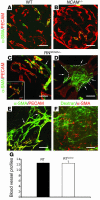
Pathological organization of periendothelial α-SMA+ cells correlates with increased tumor vessel leakage in RTNC/KO angiogenic islets. Pancreas sections from 8-week-old mice were double-immunostained with antibodies against PECAM (red) and α-SMA (AE, green; F, red). In WT (A) and NCAM–/– (B) islets, α-SMA+ cells were closely attached to the endothelium. Premature abnormal organization of periendothelial α-SMA+ cells, including detachment of α-SMA+ cells from endothelial cells (arrow in C) and multiple layers of α-SMA+ cells with an apparent loose attachment to the endothelium (D), and presence of fibroblast-like α-SMA+ cells in RTNCAM+/– angiogenic islets (E), were observed in RTNCAM+/– islets. The dashed lines and arrows in D indicate the borders of the endothelium and α-SMA+ cells stretching away from the vessel, respectively. (F) Pancreas section of an 8-week-old RTNCAM+/– mouse perfused with FITC-dextran (green), immunostained with anti–α-SMA (red). Increased leakage correlated with severely disorganized α-SMA+ periendothelial cells. The inset in C shows coexpression of NG2 (green) and α-SMA (red). (G) Analysis of blood vessel density revealed no difference between RT and RTNC/KO islets. Average values ± SEM are shown. Scale bars: 50 μm (AF).
Figure 3
Figure 3
Ectopic expression of NCAM in T241 tumor cells stimulates tumor growth and improves pericyte recruitment and coverage. (A and B) Tumor sections of T241eGFP (A) and T241NCAM (B) cells transplanted on WT mice double-stained with antibodies against PECAM (red) and α-SMA (green). (C and D) Pericyte recruitment and coverage, but not integration (C), together with the number of vessel profiles (D), increased in T241NCAM compared with T241eGFP tumors. (E and F) Independent of whether grafted on WT (E and F) or NCAM–/– (F) mice, growth of T241NCAM tumors (WT, n = 17; NCAM–/–, n = 10) was significantly increased compared with growth of T241eGFP tumors (WT, n = 20; NCAM–/–, n = 10) (F). (E) Tumor growth curves of T241eGFP and T241NCAM tumor cells grafted on WT mice (n = 6–8 for each genotype and time point). The inset shows examples of tumors at day 9. Average values ± SEM are shown. Scale bars: 50 μm (A and B) and 5 mm (E). *P < 0.05, **P < 0.01, ***P < 0.001 (2-tailed Student’s t test).
Figure 4
Figure 4
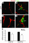
NCAM-induced improvement of pericyte recruitment and coverage requires proper PDGF-B production by host-derived tumor stroma. (AD) Tumor sections of T241eGFP (A and B) and T241NCAM (C and D) cells transplanted on Pdgfbret/ret mice double-stained with antibodies against PECAM (red) and α-SMA (green). Pericyte recruitment (A and C) and association with the endothelium (B and D) were impaired in the majority of the blood vessels, independent of whether T241eGFP or T241NCAM cells were transplanted. The arrows indicate the detachment of pericytes from the endothelium. Scale bars: 20 μm. (E) PDGF-B and PDGFR-β mRNA expression is downregulated in T241NCAM tumors compared with T241eGFP tumors (transplanted on WT mice). Average values ± SEM are shown. The mean for T241eGFP is set to 1 to indicate fold change in T241NCAM tumors. *P < 0.05, **P < 0.01 (2-tailed Student’s t test).
Figure 5
Figure 5
Pericyte detachment causes tumor cell dissemination in distant organs and local lymph nodes. (A) Intercrosses of RT and Pdgfbret/ret mice result in β tumor cell dissemination in distant organs, including the liver, kidney, and intestine (arrows), and local lymph nodes (arrowheads). (B and C) Immunostainings of sections of RT adenomas and carcinomas and RTPdgfbret/ret local lymph node and distant organ metastases with Pdx1 (B) and E-cadherin (C) antibodies. Exocrine and tumor tissues are indicated by “E” and “T,” respectively. (DG) Pancreatic tumor sections from RTNC/KO (D and F) and RTPdgfbret/ret (E and G) mice were stained with H&E. Tissue disaggregation and presence of isolated tumor cell clusters inside hemorrhagic lacunae were observed in both genotypes. Isolated cell clusters are indicated by arrows. Scale bars: 1 cm (A), 100 μm (BE), and 50 μm (F and G).
Figure 6
Figure 6
Severe α-SMA+ cellular phenotypes are associated with changes in the expression and distribution of ECM molecules. Pancreas sections of 8-week-old WT (A, D, and G), RT (B, E, and H), and RTNCAM+/– (C, F, and I) mice were double-immunostained with antibodies against α-SMA (green) and collagen IV (AC), fibronectin (DF), and laminin (GI) (red). In WT islets, α-SMA+ cells are closely attached to the blood vessel endothelium. The middle and right columns show angiogenic islets in RT (B, E, and H) and RTNCAM+/– (C, F, and I) mice. Whereas the distribution of the examined ECM molecules in RT islets was commonly observed in RTNC/KO islets, the images of consecutive sections of an RTNCAM+/– pancreas show changes in ECM deposition specifically observed in areas with fibroblast-like α-SMA+ cells in RTNC/KO islets, i.e., less deposition of both collagen IV (C) and laminin (I). The insets in C and I show normal islets from the same sections. The inset in F shows the same area stained with PECAM (red) antibodies, showing the presence of blood vessels (arrows). In contrast to laminin α1 and collagen IV, fibronectin lost its perivascular distribution and became more widely distributed (F). Scale bars: 50 μm.
Figure 7
Figure 7
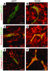
Ectopic expression of NCAM improves deposition of perivascular ECM components in T241 tumors. Sections from T241eGFP (A, C, and E) and T241NCAM (B, D, and F) tumors were stained with antibodies against laminin α1 (red; A and B), fibronectin (red; C and D), collagen IV (red; E and F), and PECAM (green; AF). The laminin α1 and fibronectin expression was upregulated both perivascularly and in the tumor tissue in T241NCAM tumors (B and D) compared with T241eGFP (A and C). Collagen IV exhibited more distinct distribution to the blood vessels in T241NCAM tumors (F) compared with T241eGFP (E). Scale bars: 20 μm.
Figure 8
Figure 8
Lymphangiogenesis is unaffected during RTNC/KO tumor progression. Immunostainings of sections of RT (left) and RTNCAM+/– (right) islets with LYVE-1 antibodies (red). Nuclei are indicated by DAPI (blue). Scale bars: 100 μm.

References

    1. Ellis LM, Fidler IJ. Angiogenesis and metastasis. Eur. J. Cancer. 1996;32A:2451–2460. - PubMed
    1. Thiery JP. Epithelial-mesenchymal transitions in tumour progression. Nat. Rev. Cancer. 2002;2:442–454. - PubMed
    1. Hanahan D. Heritable formation of pancreatic beta-cell tumours in transgenic mice expressing recombinant insulin/simian virus 40 oncogenes. Nature. 1985;315:115–122. - PubMed
    1. Perl AK, et al. Reduced expression of neural cell adhesion molecule induces metastatic dissemination of pancreatic beta tumor cells. Nat. Med. 1999;5:286–291. - PubMed
    1. Roesler J, Srivatsan E, Moatamed F, Peters J, Livingston EH. Tumor suppressor activity of neural cell adhesion molecule in colon carcinoma. Am. J. Surg. 1997;174:251–257. - PubMed

Publication types

MeSH terms

Substances