. 2006 Feb;14(2):247-55.
doi: 10.1016/j.str.2005.10.010.

Antiparallel four-stranded coiled coil specified by a 3-3-1 hydrophobic heptad repeat

Affiliations

Antiparallel four-stranded coiled coil specified by a 3-3-1 hydrophobic heptad repeat

Yiqun Deng et al. Structure. 2006 Feb.

Abstract

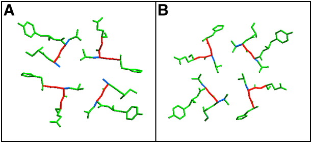
Coiled-coil sequences in proteins commonly share a seven-amino acid repeat with nonpolar side chains at the first (a) and fourth (d) positions. We investigate here the role of a 3-3-1 hydrophobic repeat containing nonpolar amino acids at the a, d, and g positions in determining the structures of coiled coils using mutants of the GCN4 leucine zipper dimerization domain. When three charged residues at the g positions in the parental sequence are replaced by nonpolar alanine or valine side chains, stable four-helix structures result. The X-ray crystal structures of the tetramers reveal antiparallel, four-stranded coiled coils in which the a, d, and g side chains interlock in a combination of knobs-into-knobs and knobs-into-holes packing. Interfacial interactions in a coiled coil can therefore be prescribed by hydrophobic-polar patterns beyond the canonical 3-4 heptad repeat. The results suggest that the conserved, charged residues at the g positions in the GCN4 leucine zipper can impart a negative design element to disfavor thermodynamically more stable, antiparallel tetramers.


Figures

Figure 1
Figure 1
Helical Wheel Representation of the GCN4-pR Leucine Zipper as a Parallel, Two-Stranded Coiled Coil The view is from the N terminus looking down the superhelical axis. Heptad repeat positions are labeled a–g. Prime (′) refers to positions from the second helix. In the mutant peptides described here, three charged residues in the dashed box at position g were changed to alanine and valine. The sequences (with the three mutated g positions underlined) are the following: GCN4-pR, MK VKQLEDK VEELLSK NYHLENE VARLKKL VGER; GCN4-pA, MK VKQLEDA VEELLSA NYHLENA VARLKKL VGER; GCN4-pV, MK VKQLEDV VEELLSV NYHLENV VARLKKL VGER. The GCN4-pR sequence contains an additional Met-Lys-Val and no Arg-Met at its N terminus, but is otherwise identical to GCN4-p1.
Figure 2
Figure 2
The GCN4-pA and GCN4-pV Peptides Form Four-Stranded Helical Bundles GCN4-pA, triangles; GCN4-pV, circles. (A) Circular dichroism (CD) spectra of 50 μM peptide at 4°C in PBS (pH 7.0). (B) Thermal melts monitored by CD at 222 nm. The squares show data for the parent GCN4-pR peptide. (C) Sedimentation equilibrium data for a 50 μM sample of GCN4-pA at 20°C and 28,000 rpm in PBS (pH 7.0). The data fit closely to a tetramer bundle. Curves expected for trimeric and pentameric models are indicated for comparison. The deviation in the data from the linear fit for a tetrameric model is plotted (upper). (D) Sedimentation equilibrium data for a 500 μM sample of GCN4-pV at 20°C and 28,000 rpm in PBS (pH 7.0).
Figure 3
Figure 3
Crystal Structure of the GCN4-pA Tetramer (A) Lateral view of the tetramer. Red van der Waals surfaces identify residues at the a positions, green surfaces identify residues at the d positions, and yellow surfaces identify residues at the g positions. The N termini of helices A and B are indicated. (B) Axial view of the tetramer. The green, yellow, and red van der Waals surfaces of the L6 (d), L30 (g), and V31 (a) side chains are depicted. (C) Cross-section of the superhelix in the L20 (d) layer. The 1.5 Å 2Fo – Fc electron density map (contoured at 1.5σ) is shown with the refined molecular model. (D) Helical wheel projection of residues 2–32 of the GCN4-pA tetramer. Heptad repeat positions are labeled a–g. The leucines at the d positions form the apolar interface of the tetramer.
Figure 4
Figure 4
Crystal Structure of the GCN4-pV Tetramer (A) Lateral view of the tetramer. Red van der Waals surfaces identify residues at the a positions, green surfaces identify residues at the d positions, and yellow surfaces identify residues at the g positions. The N termini of helices A and B are indicated. (B) Axial view of the tetramer. The green, yellow, and red van der Waals surfaces of the L6 (d), L30 (g), and V31 (a) side chains are depicted. (C) Cross-section of the superhelix in the L20 (d) layer. The 2Fo – Fc electron density map at 1.2σ contour is shown with the refined molecular model. (D) Superposition of the backbone conformations of the parallel GCN4-pIL tetramer (red) and the antiparallel GCN4-pA (green) and GCN4-pV (yellow) tetramers.
Figure 5
Figure 5
Knobs-into-Holes Packing in the Antiparallel GCN4-pA and GCN4-pV Tetramers Helix cross-sectional layers centered on the a, c, d, e, and g positions of GCN4-pA (A) and GCN4-pV (B) are depicted. Knobs formed by the side chains of one helix fit into holes formed by the spaces between side chains on a neighboring helix. The Cα-Cβ bond of each knob (thick blue line) and the Cα-Cα vector at the base of the recipient hole on the neighboring helix (thick red line) are indicated.
Figure 6
Figure 6
Core Packing in Antiparallel, Four-Stranded Coiled Coils (A–C) Helical wheel representation of the antiparallel tetramer showing the hydrophobic interface formed by side chains from positions a,d (A), a,d,e (B), and a,d,g (C). Heptad repeat positions are labeled a–g. (D) Buried surface areas in WSPLB 21–52 (a,d packing), the lac repressor tetramerization domain (a,d,e packing), rop (a,d packing), GCN4-pA (a,d,g packing), and GCN4-pV (a,d,g packing). Percent buried surface area is expressed as the fraction of accessible side chain surface area in the isolated helix that becomes buried in the antiparallel tetramer.

References

    1. Alberti S., Oehler S., von Wilcken-Bergmann B., Muller-Hill B. Genetic analysis of the leucine heptad repeats of Lac repressor: evidence for a 4-helical bundle. EMBO J. 1993;12:3227–3236. - PMC - PubMed
    1. Banner D.W., Kokkinidis M., Tsernoglou D. Structure of the ColE1 rop protein at 1.7 Å resolution. J. Mol. Biol. 1987;196:657–675. - PubMed
    1. Brunger A.T., Adams P.D., Clore G.M., DeLano W.L., Gros P., Grosse-Kunstleve R.W., Jiang J.S., Kuszewski J., Nilges M., Pannu N.S. Crystallography & NMR system: a new software suite for macromolecular structure determination. Acta Crystallogr. D Biol. Crystallogr. 1998;54:905–921. - PubMed
    1. Bryson J.W., Betz S.F., Lu H.S., Suich D.J., Zhou H.X., O'Neil K.T., DeGrado W.F. Protein design: a hierarchic approach. Science. 1995;270:935–941. - PubMed
    1. Cantor C., Schimmel P. W.H. Freeman; New York: 1980. Biophysical Chemistry, Volume III.

Publication types

MeSH terms

Substances