. 2006 Feb 21;103(8):2764-9.
doi: 10.1073/pnas.0511185103. Epub 2006 Feb 10.

An unexpected antibody response to an engineered influenza virus modifies CD8+ T cell responses

Affiliations

An unexpected antibody response to an engineered influenza virus modifies CD8+ T cell responses

Paul G Thomas et al. Proc Natl Acad Sci U S A. .

Abstract

The ovalbumin(323-339) peptide that binds H2I-A(b) was engineered into the globular heads of hemagglutinin (H) molecules from serologically non-cross-reactive H1N1 and H3N2 influenza A viruses, the aim being to analyze recall CD4+ T cell responses in a virus-induced respiratory disease. Prime/challenge experiments with these H1ova and H3ova viruses in H2(b) mice gave the predicted, ovalbumin-specific CD4+ T cell response but showed an unexpectedly enhanced, early expansion of viral epitope-specific CD8+ T cells in spleen and a greatly diminished inflammatory process in the virus-infected respiratory tract. At the same time, the primary antibody response to the H3N2 challenge virus was significantly reduced, an effect that has been associated with preexisting neutralizing antibody in other experimental systems. Analysis of serum from the H1ova-primed mice showed low-level binding to H3ova but not to the wild-type H3N2 virus. Experiments with CD4+ T cell-depleted and Ig-/- mice indicated that this cross-reactive Ig is indeed responsible for the modified pathogenesis after respiratory challenge. Furthermore, the effect does not seem to be virus-dose related, although it does require infection. These findings suggest intriguing possibilities for vaccination and, at the same time, emphasize that engineered modifications in viruses may have unintended immunological consequences.


Conflict of interest statement

Conflict of interest statement: No conflicts declared.

Figures

Fig. 1.
Fig. 1.
Model depicting the insertion of the OVA323–339 peptide (red) into the globular head of H1 (yellow). The engineered H3 is essentially identical. The insertion site is in the loop near antigenic site B. Although the structure of the loop with the OVA peptide is hypothetical, it illustrates why the change is likely to promote antibody binding. This model was generated by S. White (St. Jude Children’s Research Hospital).
Fig. 2.
Fig. 2.
The response to secondary challenge, measured for five mice per group. (A) Quantification of OT-II-specific CD4+ T cells by ELISPOT analysis of spleen on day 8. (B) Virus titers in lung as determined by Madin–Darby canine kidney (MDCK) cell plaque assay. (C) Serum antibody titers measured by antigen-specific ELISA on days 2, 5, and 8 of secondary challenge. (D) Absence of a CD8+ T cell response on day 8 after homologous challenge of H3wt-infected mice. Results are expressed as mean ± SE (n = 5); ∗, P < 0.05.
Fig. 3.
Fig. 3.
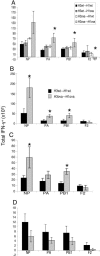
Measuring epitope-specific CD8+ T cell responses in spleen on day 2 (A), day 5 (B), and day 8 (C) and bronchoalveolar lavage (BAL) on day 5 (D) for the H3wt→H1wt and H3ova→H1ova challenges. Influenza virus epitopes are as follows: NP, nucleoprotein DbNP366-374; PA and PB1, RNA polymerase subunits PA (DbPA224-233) and PB1 (KbPB1703-711); F2, an alternatively spliced PB1 (DbPB1-F262-70). Results were determined by intracellular cytokine (IFN-γ) staining and are expressed as mean ± SE (n = 5); ∗, P < 0.05. Experiments on each day were repeated at least twice.
Fig. 4.
Fig. 4.
Epitope-specific CD8+ T cell responses in spleen (A and B) and BAL (C and D) after high-dose [106 egg 50% infectious dose (EID50)] (A and C) or low-dose (104 EID50) (B and D) secondary intranasal (i.n.) challenge. NS2, nonstructural protein 2. Representative data from two independent experiments are shown. Results were determined by intracellular cytokine staining on day 8 after challenge and are expressed as mean ± SE (n = 5); ∗, P < 0.05.
Fig. 5.
Fig. 5.
Splenic enhancement phenotype is dependent on a primed B-cell response. (A) The enhanced spleen response is seen only for the H3ova→H1ova combination. H3ova→H1wt and H3wt→H1ova infections produce responses equivalent to H3wt→H1wt. (B and C) Depleting the CD4+ T cells by treatment with the GK1.5 monoclonal antibody (C) every second day from day 3 before secondary challenge (30) causes some decrease in the overall magnitude of the response vs. undepleted mice (B) but does not change the enhancement characteristic of the H3ova→H1ova challenge. These data are representative of three independent experiments. The percentage of CD4+ T cells found by flow cytometric analysis of spleen was always <2% in the depleted mice. (D) The response in Ig−/− μMT (B cell knockout) mice after secondary challenge with the indicated viruses. All results were measured by intracellular cytokine staining on day 5 and are expressed as mean ± SE (n = 5); ∗, P < 0.05.

References

    1. Stephenson J. R. Curr. Pharm. Biotechnol. 2001;2:47–76. - PubMed
    1. Belz G. T., Xie W., Doherty P. C. J. Immunol. 2001;166:4627–4633. - PubMed
    1. Riberdy J. M., Christensen J. P., Branum K., Doherty P. C. J. Virol. 2000;74:9762–9765. - PMC - PubMed
    1. Luo G., Chung J., Palese P. Virus Res. 1993;29:141–153. - PubMed
    1. Both G. W., Sleigh M. J., Cox N. J., Kendal A. P. J. Virol. 1983;48:52–60. - PMC - PubMed

Publication types

MeSH terms