. 2006 Feb 21;103(8):2677-82.
doi: 10.1073/pnas.0511084103. Epub 2006 Feb 10.

Z-DNA-forming sequences generate large-scale deletions in mammalian cells

Affiliations

Z-DNA-forming sequences generate large-scale deletions in mammalian cells

Guliang Wang et al. Proc Natl Acad Sci U S A. .

Abstract

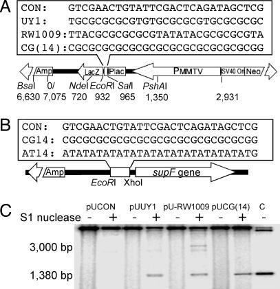
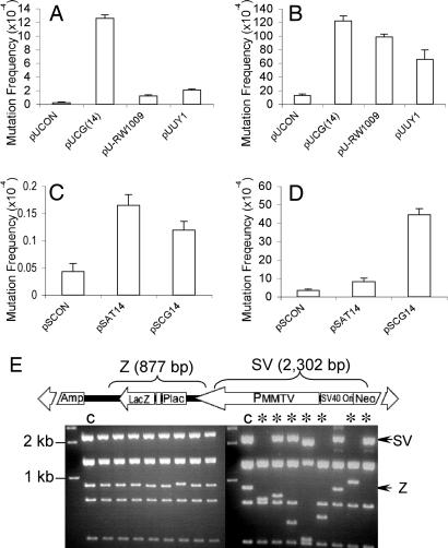
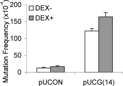
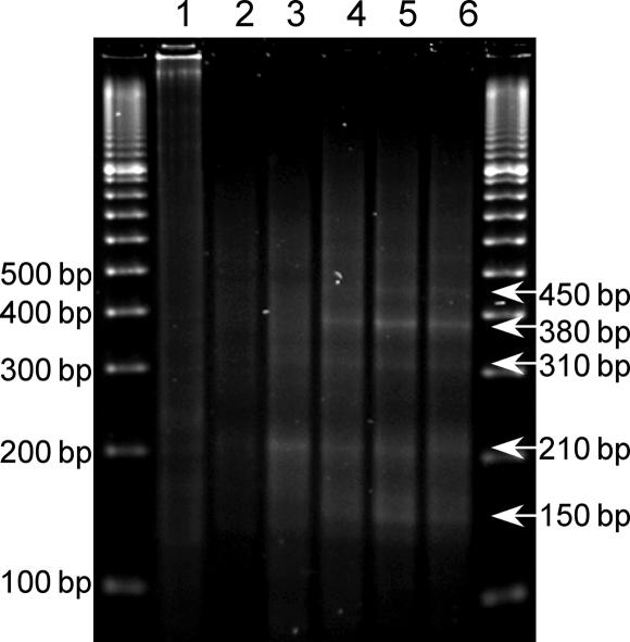
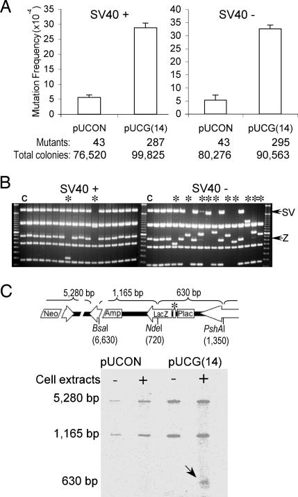
Spontaneous chromosomal breakages frequently occur at genomic hot spots in the absence of DNA damage and can result in translocation-related human disease. Chromosomal breakpoints are often mapped near purine-pyrimidine Z-DNA-forming sequences in human tumors. However, it is not known whether Z-DNA plays a role in the generation of these chromosomal breakages. Here, we show that Z-DNA-forming sequences induce high levels of genetic instability in both bacterial and mammalian cells. In mammalian cells, the Z-DNA-forming sequences induce double-strand breaks nearby, resulting in large-scale deletions in 95% of the mutants. These Z-DNA-induced double-strand breaks in mammalian cells are not confined to a specific sequence but rather are dispersed over a 400-bp region, consistent with chromosomal breakpoints in human diseases. This observation is in contrast to the mutations generated in Escherichia coli that are predominantly small deletions within the repeats. We found that the frequency of small deletions is increased by replication in mammalian cell extracts. Surprisingly, the large-scale deletions generated in mammalian cells are, at least in part, replication-independent and are likely initiated by repair processing cleavages surrounding the Z-DNA-forming sequence. These results reveal that mammalian cells process Z-DNA-forming sequences in a strikingly different fashion from that used by bacteria. Our data suggest that Z-DNA-forming sequences may be causative factors for gene translocations found in leukemias and lymphomas and that certain cellular conditions such as active transcription may increase the risk of Z-DNA-related genetic instability.


Conflict of interest statement

Conflict of interest statement: No conflicts declared.

Figures

Fig. 1.
Fig. 1.
Z-DNA structures formed at the inserts. (A and B) Schematic structure of the mutation reporter shuttle plasmid, pUCNIM (A) and pSP189 (B). (Upper) Positions and sequences of inserts are shown. (Lower) The diagnostic restriction sites used in this study are listed. (C) Identification of the Z-DNA conformation at the insertion site. The insert-containing plasmids were treated with single-stranded DNA-specific S1 nuclease followed by BsaI digestion. The ≈3,000-bp fragment and a shorter ≈1,380-bp fragment released by S1 nuclease cleavage from pUCON and Z-DNA forming plasmids, respectively, are indicated. Lane C is a 1,377-bp BsaI–EcoRI fragment as a size standard.
Fig. 2.
Fig. 2.
Z-DNA-induced mutations in bacterial cells and mammalian COS-7 cells. (A) Z-DNA-induced lacZ′ mutation frequencies in DH5α cells. (B) Z-DNA-induced lacZ′ mutation frequencies in mammalian COS-7 cells. (C) Z-DNA-induced supF mutation frequencies in DH5α cells. (D) Z-DNA-induced supF mutation frequencies in mammalian COS-7 cells. A total of >100,000 colonies were counted in each group. Error bars show the SEM of three or four independent experiments. (E) pUCG(14) mutants derived from DH5α cells (Left) and COS-7 cells (Right) were digested with EagI and BssSI, resulting in seven fragments. The 877-bp fragment containing the Z-DNA-forming sequence (Z) and the 2,302-bp fragment between the Z-DNA locus and SV40 Ori (SV) are shown schematically (Upper) and marked with arrows in the gel. ∗ indicates large-scale deletions (≥50 bp); C refers to control sample.
Fig. 3.
Fig. 3.
Transcription increases both spontaneous and Z-DNA-induced mutagenesis. After transfection with pUCON or pUCG(14) plasmids, COS-7 cells were incubated for 48 h in the absence or presence of 10 μM dexamethasone. Mutants in the recovered plasmids were screened. A total of >200,000 colonies were counted in each group. Error bars show the SEM of three or four independent experiments.
Fig. 4.
Fig. 4.
LM-PCR analysis of Z-DNA-induced DSBs. LM-PCR products were subjected to agarose gel electrophoresis and visualized by ethidium bromide staining. Lane 1, pUCG(14) prepared from DH5α cells; lane 2, pUCON replicated in COS-7 cells for 48 h; lanes 3–6, pUCG(14) replicated in COS-7 cells for 4, 8, 24, and 48 h, respectively. Sizes of detected bands are listed on the right.
Fig. 5.
Fig. 5.
Replication-dependent and independent Z-DNA-induced genetic instability in mammalian cell extracts. (A) Replication-proficient (Left) and replication-deficient (Right) Z-DNA-induced mutagenesis in HeLa cell extracts. The number of mutants observed and the total number of colonies screened are listed below the corresponding bars. Error bars show the SEM of three independent experiments. (B) Restriction analysis of mutants generated in replicated (Left) and unreplicated (Right) systems. ∗ refers to large-scale deletions (≥50 bp); C refers to control sample. (C) Z-DNA-induced cleavages in HeLa cell extracts in the absence of replication. The positions of three restriction sites on the plasmid, pUCNIM, are shown schematically above. The Z-DNA locus is marked with ∗. A radiolabeled 630-bp fragment containing breakages generated in HeLa cell extracts is indicated by an arrow.

References

    1. Wang G., Vasquez K. M. Proc. Natl. Acad. Sci. USA. 2004;101:13448–13453. - PMC - PubMed
    1. Adachi M., Tsujimoto Y. Oncogene. 1990;5:1653–1657. - PubMed
    1. Seite P., Leroux D., Hillion J., Monteil M., Berger R., Mathieu-Mahul D., Larsen C. J. Genes Chromosomes Cancer. 1993;6:39–44. - PubMed
    1. Wolfl S., Wittig B., Rich A. Biochim. Biophys. Acta. 1995;1264:294–302. - PubMed
    1. Rimokh R., Rouault J. P., Wahbi K., Gadoux M., Lafage M., Archimbaud E., Charrin C., Gentilhomme O., Germain D., Samarut J., et al. Genes Chromosomes Cancer. 1991;3:24–36. - PubMed

Publication types