. 2006 Feb 21;103(8):2512-7.
doi: 10.1073/pnas.0506020102. Epub 2006 Feb 10.

A macroporous hydrogel for the coculture of neural progenitor and endothelial cells to form functional vascular networks in vivo

Affiliations

A macroporous hydrogel for the coculture of neural progenitor and endothelial cells to form functional vascular networks in vivo

Millicent C Ford et al. Proc Natl Acad Sci U S A. .

Abstract

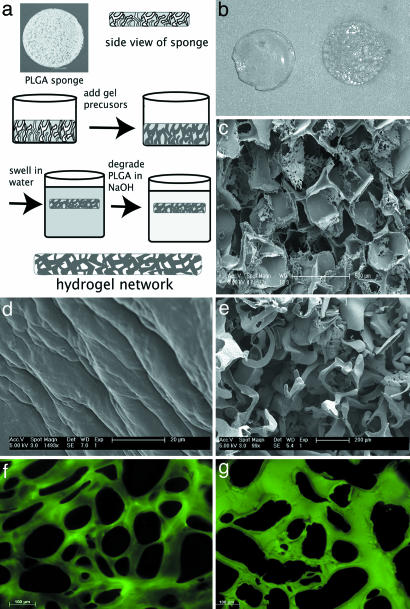
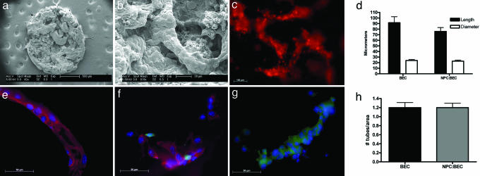
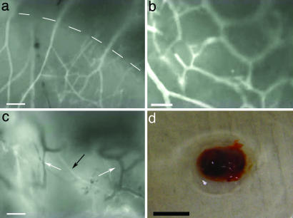
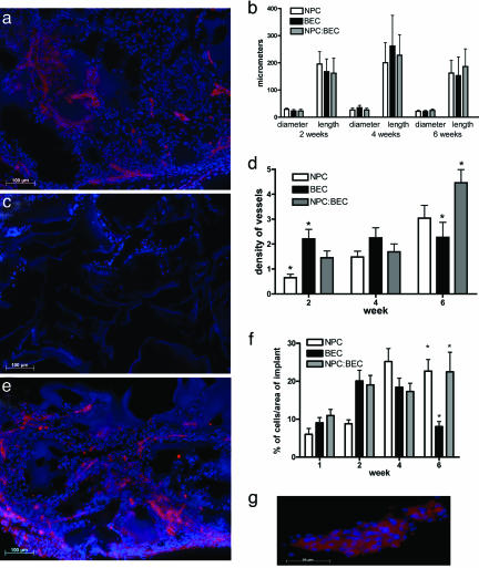
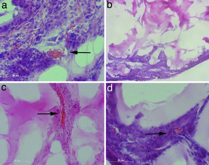
A microvascular network is critical for the survival and function of most tissues. We have investigated the potential of neural progenitor cells to augment the formation and stabilization of microvascular networks in a previously uncharacterized three-dimensional macroporous hydrogel and the ability of this engineered system to develop a functional microcirculation in vivo. The hydrogel is synthesized by cross-linking polyethylene glycol with polylysine around a salt-leached polylactic-co-glycolic acid scaffold that is degraded in a sodium hydroxide solution. An open macroporous network is formed that supports the efficient formation of tubular structures by brain endothelial cells. After subcutaneous implantation of hydrogel cocultures in mice, blood flow in new microvessels was apparent at 2 weeks with perfused networks established on the surface of implants at 6 weeks. Compared to endothelial cells cultured alone, cocultures of endothelial cells and neural progenitor cells had a significantly greater density of tubular structures positive for platelet endothelial cell adhesion molecule-1 at the 6-week time point. In implant cross sections, the presence of red blood cells in vessel lumens confirmed a functional microcirculation. These findings indicate that neural progenitor cells promote the formation of endothelial cell tubes in coculture and the development of a functional microcirculation in vivo. We demonstrate a previously undescribed strategy for creating stable microvascular networks to support engineered tissues of desired parenchymal cell origin.


Conflict of interest statement

Conflict of interest statement: No conflicts declared.

Figures

Fig. 1.
Fig. 1.
Formation of the macroporous hydrogel. (a) Schematic of the macroporous process. (b) Photograph of isotropic hydrogel (Left) and macroporous hydrogel (Right). The gels are 5 mm in diameter. (c) SEM micrograph of a salt-leached scaffold used to create the macroporous architecture. (d and e) SEM micrographs of an isotropic hydrogel (d) and a macroporous hydrogel (e). The macropores, created by casting the hydrogel around the salt-leached scaffold, are evident. (f and g) Cross sections of the isotropic (f) and macroporous (g) hydrogel labeled with FITC to demonstrate the pore structure of the gels. The FITC labels the amines in the polylysine component of the hydrogel.
Fig. 2.
Fig. 2.
In vitro study after 3 days in culture. (a) Scanning electron micrograph of BECs cultured on PEG-polylysine hydrogel at low magnification. The entire 5-mm disk is visible and uniformly seeded with BECs. (b) SEM micrograph of the hydrogel at higher magnification, demonstrating BEC tubule formation. (c) Live image of DiI-stained BECs following the porous architecture of the hydrogel. (d) Graph of BEC tubule length and diameter. There are no significant differences between the treatment groups BEC and NPC:BEC. Error bars ± SE. (eg) Immunocytochemical staining of fixed hydrogels from the three treatment groups. BECs are stained for PECAM-1(red) and DAPI (blue). NPCs are green because of GFP expression. (e) BEC hydrogel. (f) NPC:BEC hydrogel. (g) NPC hydrogel. (h) Graph of the number of tubules per area at ×10 magnification. There are no significant differences between groups BEC and NPC:BEC. Error bars ± SE.
Fig. 3.
Fig. 3.
Intravital video images of blood flow after retro-orbital injection of FITC-dextran. (a) Six-week NPC:BEC hydrogel. Dotted line demarcates the hydrogel/host interface. (Scale bar: 100 μm.) (b) Six-week NPC:BEC hydrogel. (Scale bar: 50 μm.) (c) Four-week BEC hydrogel. White arrows point to vessels lacking FITC-dextran, and the black arrow points to vessels with FITC-dextran. (Scale bar: 100 μm.) (d) Clot retrieved from BEC hydrogel after 6 weeks in vivo. (Scale bar: 5 mm.)
Fig. 4.
Fig. 4.
In vivo immunohistochemical staining and quantification. (a) Six-week in vivo NPC hydrogel stained for PECAM-1 (red) and DAPI (blue). At 6 weeks, host vessels are seen in the implant. (b) The graph of the dimensions shows that there are no significant differences over any of the time points for any of the groups with regard to the average length or diameter of the tubules. Error bars ± SE. (c) Six-week BEC hydrogel stained for PECAM-1 and DAPI. The central region of the gel is devoid of cells or vessels. (d) The density graph shows that although the density of vessels is constant for the BEC group, the density is increasing for the NPC:BEC group over the time points studied. Furthermore, there is a significantly greater (P < 0.05) density of vessels in the NPC:BEC group as compared with the BEC group at 6 weeks. Error bars ± SE. (e) Six-week NPC:BEC hydrogel stained for PECAM-1 and DAPI. There are a large number of vessels throughout the gel. (f) Graph of the percentage of cells over the area of the implant. After 6 weeks in vivo, NPC and NPC:BEC hydrogels have a greater (P < 0.05) percentage of cells than the BEC implants. Moreover, there is a significant increase in the percentage of cells from 1 to 6 weeks for NPC and NPC:BEC groups. Error bars ± SE. (g) High-magnification PECAM-positive vessel from NPC:BEC implants after 6 weeks in vivo.
Fig. 5.
Fig. 5.
Hematoxylin and eosin staining of in vivo implants. (a) Four-week BEC hydrogel. Red blood cells are evident in the vessel lumen (b) Six-week BEC hydrogel. Few cells or vessels are found at the center of the implant. (c) Four-week NPC:BEC hydrogel. (d) Six-week BEC:NPC hydrogel. Red blood cells are seen at 4 and 6 weeks in NPC:BEC hydrogels. Arrows denote red blood cells in the blood vessel lumen.

References

    1. Peters M. C., Polverini P. J., Mooney D. J. J. Biomed. Mate. Res. 2002;60:668–678. - PubMed
    1. Smith M. K., Peters M. C., Richardson T. P., Garbern J. C., Mooney D. J. Tissue Eng. 2004;10:63–71. - PubMed
    1. Takeshita S., Zheng L. P., Brogi E., Kearney M., Pu L. Q., Bunting S., Ferrara N., Symes J. F., Isner J. M. J. Clin. Invest. 1994;93:662–670. - PMC - PubMed
    1. Richardson T. P., Peters M. C., Ennett A. B., Mooney D. J. Nat. Biotechnol. 2001;19:1029–1034. - PubMed
    1. Lee H., Cusick R. A., Browne F., Kim T. H., Ma P. X., Utsunomiya H., Langer R., Vacanti J. P. Transplantation. 2002;73:1589–1593. - PubMed

Publication types