. 2006 Mar;18(3):545-59.
doi: 10.1105/tpc.105.039156. Epub 2006 Feb 10.

The Arabidopsis-mei2-like genes play a role in meiosis and vegetative growth in Arabidopsis

Affiliations

The Arabidopsis-mei2-like genes play a role in meiosis and vegetative growth in Arabidopsis

Jagreet Kaur et al. Plant Cell. 2006 Mar.

Abstract

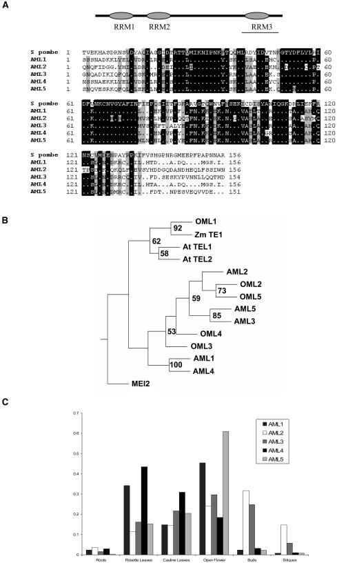
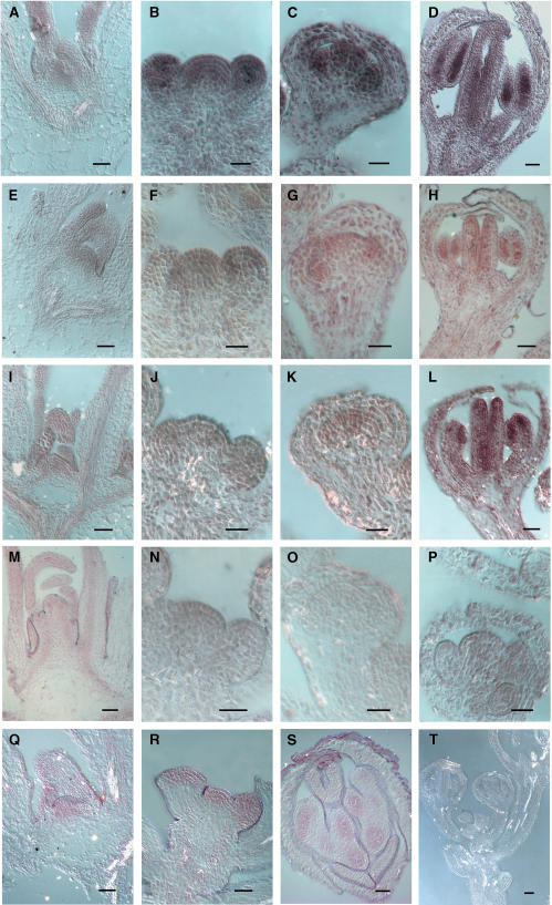
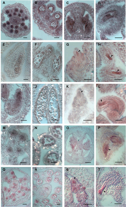
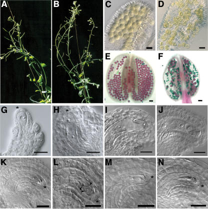
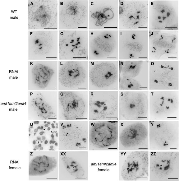
The Arabidopsis-mei2-Like (AML) genes comprise a five-member gene family related to the mei2 gene, which is a master regulator of meiosis in Schizosaccharomyces pombe and encodes an RNA binding protein. We have analyzed the AML genes to assess their role in plant meiosis and development. All five AML genes were expressed in both vegetative and reproductive tissues. Analysis of AML1-AML5 expression at the cellular level indicated a closely similar expression pattern. In the inflorescence, expression was concentrated in the shoot apical meristem, young buds, and reproductive organ primordia. Within the reproductive organs, strong expression was observed in meiocytes and developing gametes. Functional analysis using RNA interference (RNAi) and combinations of insertion alleles revealed a role for the AML genes in meiosis, with RNAi lines and specific multiple mutant combinations displaying sterility and a range of defects in meiotic chromosome behavior. Defects in seedling growth were also observed at low penetrance. These results indicate that the AML genes play a role in meiosis as well as in vegetative growth and reveal conservation in the genetic mechanisms controlling meiosis in yeast and plants.


Figures

Figure 1.
Figure 1.
AML Genes Are Expressed in Vegetative and Reproductive Tissues in Arabidopsis. (A) AML genes encode RNA binding proteins with three RRMs. Bar indicates the region of greatest similarity. ClustalX alignment of AML1-AML5 and S. pombe mei2 proteins for RRM3. (B) Phylogenetic tree of mei2-related genes from Arabidosis, rice, maize, and S. pombe generated using Phylip. Bootstrap values >50 are indicated. (C) RT-PCR analysis of relative expression of AML1-AML5 in different tissues normalized with GAPC.
Figure 2.
Figure 2.
RNA in Situ Expression Analysis of AML1-AML5 in Apical Meristems and Reproductive Organs. AML5 ([A] to [D]); AML1 ([E] to [H]); AML2 ([I] to [L]); AML3 ([M] to [P]); AML4 ([Q] to [S]); AML1 sense (T). Bars = 10 μm. (A), (E), (I), (M), and (Q) Vegetative SAM. (B), (F), (J), (N), and (R) Inflorescence SAM. (C), (G), (K), and (O) Floral stage 5 and 6 buds showing increased signal in stamen and pistil primordia. (D), (H), (L), and (S) Concentrated signal is detected in anther locules and inner margins of the pistil in stage 8 and 9 buds. (P) Floral stage 7.
Figure 3.
Figure 3.
RNA in Situ Expression Analysis of AML1-AML5 in Meiocytes and Gametophytes. AML5 ([A] to [D]); AML1 ([E] to [H]); AML2 ([I] to [L]); AML3 ([M] to [P]); AML4 ([Q] to [T]). Bars = 25 μm. (A), (E), (I), (M), and (Q) Transverse ([A], [M], and [Q]) and longitudinal ([E] and [I]) sections of anthers showing strong expression in pollen mother cells. (B), (F), (J), (N), and (R) Expression in developing pollen. (C), (G), (K), (O), and (S) Expression in young ovules, including the megaspore mother cell (arrowhead). (D), (H), (L), (P), and (T) Mature ovules showing strong expression in the embryo sac (arrowhead).
Figure 4.
Figure 4.
AML5-RNAi Lines Display Sterility and Defective Gametogenesis. (A) Wild-type fertile plant with elongated siliques. (B) AML5-RNAi line showing strong sterility. The siliques have failed to elongate. (C) and (D) Optical sections of cleared pollen from the wild type (C) and a strong AML5-RNAi line (D). Majority of the pollen in the RNAi anther are shriveled. (E) and (F) Alexander staining of anthers showing viable pollen (purple) in the wild-type anthers (E) compared with a majority of nonviable pollen (green) in the AML5-RNAi line (F). (G) to (N) Different stages of female gametophyte development in the wild type ([G] to [K]) and AML5-RNAi ([L] to [N]). (G) Ovule stage 2-1, wherein the megaspore mother cell has differentiated. (H) and (I) Two-nuclear ([H]; ovule stage 3-2) and four-nuclear ([I]; ovule stage 3-4) embryo sac. (J) Ovule stage 3-6 showing a mature embryo sac. (K) Stage 3-6 ovule showing an embryo sac arrested at the uninucleate stage. (L) Two enlarged cells (arrows) separated by a cell wall (arrowhead) in place of a mature embryo sac. (M) Arrested two-nuclear embryo sac. (N) Degenerating embryo sac based on the presence of optically dense material. Asterisks mark the apical epidermis below which the megaspore mother cell differentiates. Bars = 25 μm.
Figure 5.
Figure 5.
AML1-AML5 Transcript Levels Are Reduced in AML5-RNAi Lines Showing Strong Sterility. Quantitation of AML1-AML5 transcript levels in five independent T1 lines and one T2 line (R25.8) showing strong sterility. The levels of each transcript are represented as percentages normalized against the levels of AML1-AML5 gene expression observed in control plants. In each line showing strong sterility, at least three members of the AML family show a more than twofold reduction in the transcript level. The control values represent the mean for two plants: C3 and C21, representing two control vector transformants. R20 is an RNAi line that did not show any sterility.
Figure 6.
Figure 6.
Characterization of AML Expression in Lines Carrying T-DNA Insertions in the AML1-AML5 Genes. Line diagram showing the site of T-DNA insertion in each of the AML genes together with RT-PCR analysis of AML1-AML5 expression in the insertion mutants. Exons are indicated as black boxes and the untranslated regions as white boxes. Inverted triangles represent the site of T-DNA insertion in each line. The arrows represent the LB1 primers, and the short arrows represent the gene-specific primers used. The line below the gene represents the cDNA sequence amplified using gene-specific primers. Lane 1: PCR done using left border primer (LB1) in combination with a gene-specific primer. For AML1, the gene-specific primer is downstream of the site of insertion, whereas for all others, the gene-specific primer is upstream of the site of insertion. For AML1, AML2, and AML5, no amplification is seen. For AML3 and AML4, the gene and T-DNA left border junction can be identified, indicating the presence of T-DNA in the processed transcript. Lane 2: A pair of gene-specific primers was used to detect the presence of the transcripts in the insertion mutants. In aml2, aml3, and aml5 insertion mutants, the transcripts downstream of the site of insertion could not be detected. Lane 3: The primer pairs used in lane 2 were used to amplify corresponding transcripts from wild-type plants. Lanes 4 and 5: GAPC controls for mutant and wild type, respectively.
Figure 7.
Figure 7.
Seedling Arrest in aml Mutant Combinations and RNAi Lines. (A) aml1 aml4 double mutant showing seedling arrest: wild-type seedling (black arrowhead) and arrested seedling (white arrowhead). (B) Defective root growth. (C) ProAML2:β-glucuronidase expression in root tips. (D) to (H) Scanning electron microscopy of 10-d-old seedlings: the wild type ([D] and [F]), aml1 aml2 aml4 ([E] and [G]), and AML5 RNAi (H). (D) Low magnification; cotyledons have been removed. Leaves 1 and 2 have expanded. Primordia of leaves 3 and 4 are visible. (E) Whole seedling. Leaves fail to expand. (F) High magnification; cotyledons and leaves 1, 2, and 4 have been removed. Primordia of leaves 5 and 6 are visible. (G) Primordia of leaves 3 and 4 are indicated. (H) Cotyledons have been removed. Primordia of leaves 1 and 2 are indicated. Bars = 100 μm in (D) and (E) and 30 μm in (F) to (H).
Figure 8.
Figure 8.
Defective Gametogenesis in aml1 aml2 aml4 Triple Mutants. (A) Alexander staining of anther showing viable (purple) and nonviable (green) pollen. (B) Stage 3-6 ovule with embryo sac arrested at the functional megaspore stage. (C) Two cells (arrows) present along with degenerating megaspores in place of a mature embryo sac. Arrowhead marks the cell wall. (D) Degenerating embryo sac. Bars = 20 μm.
Figure 9.
Figure 9.
Defective Meiotic Chromosome Organization in AML5-RNAi and aml1 aml2 aml4 Plants. (A) Leptotene (B) Zygotene. (C) Pachytene. (D) Late diplotene. (E) Diakinesis. (F) Metaphase I. (G) Anaphase I. (H) Metaphase II showing organelle band. (I) Anaphase II. (J) Telophase II. (K) Partial desynapsis resulting in a mix of univalents and bivalents at diplotene. (L) Chromosome bridge (arrowhead) between nonhomologous chromosomes at diakinesis. (M) Clumping of chromosomes. (N) Acentric chromosome in addition to five condensed bivalents at metaphase I. (O) Chromosome bridge (arrowhead) and laggard chromosome at telophase II. (P) Univalents and chromosome fragments corresponding to pachytene stage. (Q) Disorganized diplotene stage. (R) Chromosome bridge (arrowhead) at diakinesis. (S) Clumped chromosomes at metaphase I. (T) Two univalents and acentric chromosome at diakinesis. (U) Anaphase I bridges (arrowheads). (V) Laggard chromosomes at telophase II. (W) Ten univalents. (X) Clumped chromosomes at prometaphase I. (Y) Telophase I showing absence of organelle band. (Z) Ten univalents. (XX) Chromosome bridge (arrowhead). (YY) and (ZZ) Chromosome bridges (arrowheads) at diakinesis. Bars = 10 μm.

References

    1. Agashe, B., Prasad, C.K., and Siddiqi, I. (2002). Identification and analysis of DYAD: A gene required for meiotic chromosome organisation and female meiotic progression in Arabidopsis. Development 129 3935–3943. - PubMed
    1. Alexander, M.P. (1969). Differential staining of aborted and nonaborted pollen. Stain Technol. 44 117–122. - PubMed
    1. Alonso, J.M., et al. (2003). Genome-wide insertional mutagenesis of Arabidopsis thaliana. Science 301 653–657. - PubMed
    1. Anderson, G.H., Alvarez, N.D., Gilman, C., Jeffares, D.C., Trainor, V.C., Hanson, M.R., and Veit, B. (2004). Diversification of genes encoding mei2-like RNA binding proteins in plants. Plant Mol. Biol. 54 653–670. - PubMed
    1. Anderson, G.H., and Hanson, M.R. (2005). The Arabidopsis Mei2 homologue AML1 binds AtRaptor1B, the plant homologue of a major regulator of eukaryotic cell growth. BMC Plant Biol. 5 2. - PMC - PubMed

Publication types

MeSH terms