Comparative Study
. 2006 Jun;34(6):766-74.
doi: 10.1165/rcmb.2005-0246OC. Epub 2006 Feb 10.

Role of granulocyte macrophage colony-stimulating factor during gram-negative lung infection with Pseudomonas aeruginosa

Affiliations
Comparative Study

Role of granulocyte macrophage colony-stimulating factor during gram-negative lung infection with Pseudomonas aeruginosa

Megan N Ballinger et al. Am J Respir Cell Mol Biol. 2006 Jun.

Abstract

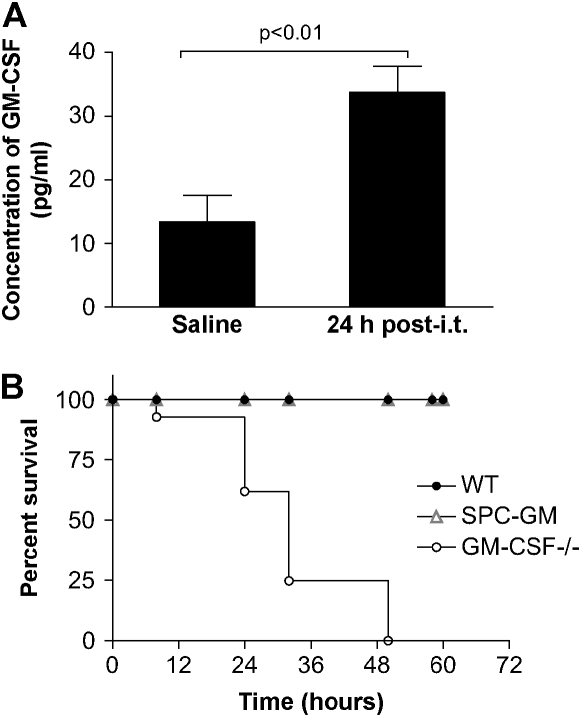
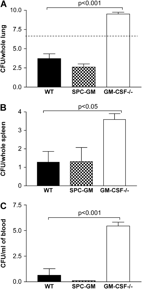
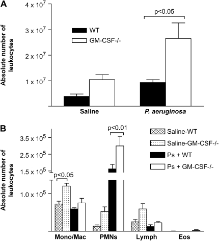
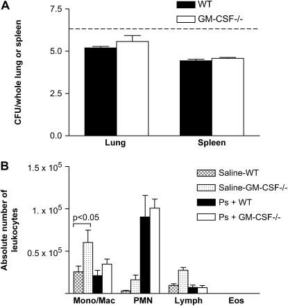
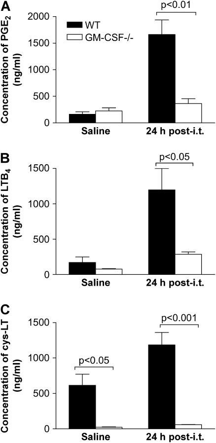
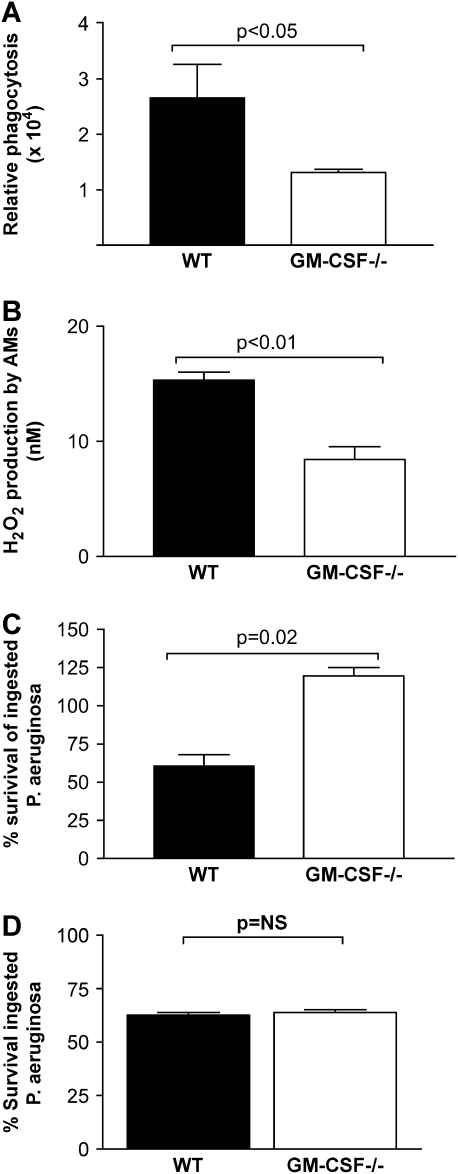
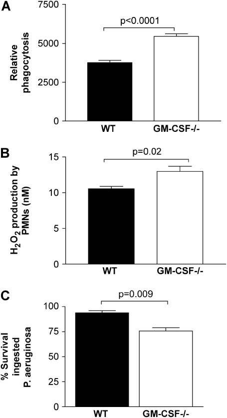
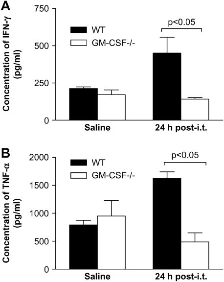
Granulocyte macrophage colony-stimulating factor (GM-CSF) stimulates survival, proliferation, differentiation, and function of myeloid cells. Recently, GM-CSF has been shown to be important for normal pulmonary homeostasis. We report that GM-CSF is induced in lung leukocytes during infection with Gram-negative bacteria. Therefore, we postulated that deficiencies in GM-CSF would increase susceptibility to Gram-negative infection in vivo. After an intratracheal inoculum with Pseudomonas aeruginosa, GM-CSF-/- mice show decreased survival compared with wild-type mice. GM-CSF-/- mice show increased lung, spleen, and blood bacterial CFU. GM-CSF-/- mice are defective in the production of cysteinyl leukotrienes, prostaglandin E2, macrophage inflammatory protein, and keratinocyte-derived chemokine in lung leukocytes postinfection. Despite these defects, inflammatory cell recruitment is not diminished at 6 or 24 h postinfection, and the functional activity of polymorphonuclear leukocytes from the lung and peritoneum against P. aeruginosa is enhanced in GM-CSF-/- mice. In contrast, alveolar macrophage (AM) phagocytosis, killing, and H2O2 production are defective in GM-CSF-/- mice. Although the absence of GM-CSF has profound effects on AMs, peritoneal macrophages seem to have normal bactericidal activities in GM-CSF-/- mice. Defects in AM function may be related to diminished levels of IFN-gamma and TNF-alpha postinfection. Thus, GM-CSF-/- mice are more susceptible to lung infection with P. aeruginosa as a result of impaired AM function.


Figures

<b>Figure 1.</b>
Figure 1.
GM-CSF is an important regulator of the innate immune response during a P. aeruginosa infection. (A) GM-CSF−/− and WT mice were inoculated intratracheally with saline or P. aeruginosa (6.8 CFU log10 scale), and lung leukocytes were isolated. We verified that GM-CSF−/− mice were not able to produce GM-CSF even when infected with P. aeruginosa, compared with an increase in the production of GM-CSF post-challenge by the WT lung leukocytes (closed bars). Data represent n = 4 per group and are representative of three similar experiments. (B) GM-CSF−/−, SPC-GM, and WT mice were infected intratracheally with P. aeruginosa at a dose of 6.0 CFU log10 scale, and survival was monitored for 72 h. At 48 h after intratracheal inoculation, the GM-CSF−/− mice (open circles) have 100% mortality compared with 100% survival of SPC-GM (gray triangles) and WT mice (closed circles). The GM-CSF−/− mice have increased mortality to P. aeruginosa infections (P = 0.0001). Data represent n = 7 mice per group and are representative of two similar experiments.
<b>Figure 2.</b>
Figure 2.
GM-CSF−/− mice have increased bacterial burden in the lungs and increased bacterial dissemination into the blood and spleen compared with SPC-GM and WT mice. Lungs, spleen, and blood were harvested 24 h after intratracheal infection with P. aeruginosa (6.7 CFU log10 scale) from GM-CSF−/−, SPC-GM, and WT mice and plated for CFU analysis. (A) There were increased CFU in the lungs of GM-CSF−/− mice (open bar) compared with the WT (closed bar) and SPC-GM mice (checkered bar) 24 h postinfection (P < 0.001). GM-CSF−/− mice (open bar) have increased bacterial dissemination as shown by higher CFU found in the spleen (B) and blood (C) compared with WT (closed bars) and SPC-GM (checkered bars) mice. Data represent n = 5 mice per group and are representative of two similar experiments. The dotted line represents the original inoculum dose (6.7 CFU log10 scale). The GM-CSF−/− mice had increased bacterial burden in their lung (P < 0.001) and increased dissemination into the spleen (P < 0.05) and blood (P < 0.001).
<b>Figure 3.</b>
Figure 3.
GM-CSF−/− mice show no defect in cellular accumulation at 24 h after P. aeruginosa. WT mice and GM-CSF−/− mice were given an intratracheal infection with P. aeruginosa at a dose of 6.5 CFU in log10 scale. Total primary lung leukocytes and inflammatory subsets were enumerated 24 h after intratracheal inoculation. (A) Total leukocytes accumulated in response to a saline treatment were not statistically different in GM-CSF−/− mice (open bars) versus WT mice (closed bars), although the trend was for the GM-CSF−/− mice to have more cells. There was a significant increase in the number of lung leukocytes in the GM-CSF−/− mice (open bars) compared with WT mice (closed bars) after P. aeruginosa (P < 0.05). (B) After saline treatment, there were more monocyte/macrophages in the GM-CSF−/− mice (dotted bars) compared with the WT mice (checked bars) (P < 0.05). In response to P. aeruginosa, PMNs are the predominant cell type in WT (closed bars) and GM-CSF−/− mice (open bars), and GM-CSF−/− mice recruit more PMNs (P < 0.01). Data represent n = 4 mice per condition and are representative of two experiments.
<b>Figure 4.</b>
Figure 4.
GM-CSF−/− mice had an intact early response against P. aeruginosa compared with WT mice. GM-CSF−/− and WT mice were infected with P. aeruginosa (6.6 CFU in log10 scale) and examined 6 h after infection. (A) The bacterial burden in the lungs and spleen of WT (closed bars) and GM-CSF−/− (open bars) mice was examined by CFU analysis. Six hours postinfection, we show no significant increase in the amount of bacterial burden in the lung or in the amount of bacterial dissemination in the spleen in GM-CSF−/− mice compared with WT mice. The dotted line represents the original inoculum dose. (B) There is no defect in the ability of GM-CSF−/− mice to recruit inflammatory subsets to the site of infection. The absolute number of monocytes/macrophages and PMNs in the GM-CSF−/− mice (open bars) compared with the WT mice (closed bars) 6 h after P. aeruginosa infection are not different. Data represent n = 5 mice per condition and are representative of two experiments.
<b>Figure 5.</b>
Figure 5.
GM-CSF−/− lung leukocytes were not able to produce eicosanoids in response to a 24 h P. aeruginosa infection. GM-CSF−/− and WT mice were inoculated with saline or P. aeruginosa (6.5 CFU in log10 scale), and 24 h later lung leukocytes were collected and incubated overnight. (A) GM-CSF−/− lung leukocytes (open bars) produced less PGE2 compared with WT (closed bars) cells. (B and C) Lung leukocytes from GM-CSF−/− mice (open bars) were unable to produce LTB4 (B) or cys-LT (C) in response to P. aeruginosa infection compared with WT lung leukocytes (closed bars). Data represent n = 5 mice per condition and are representative of two experiments.
<b>Figure 6.</b>
Figure 6.
GM-CSF−/− AMs, but not PMs, show defective phagocytosis and killing of Gram-negative bacteria in vitro. AMs were tested from WT or GM-CSF−/− mice and assayed for their ability to phagocytose or kill Gram-negative bacteria in vitro. (A) The AMs from GM-CSF−/− mice (open bars) were deficient in their ability to phagocytose FITC-labeled, Gram-negative E. coli in vitro compared with AMs from WT mice (closed bars) (P < 0.05). This assay measures only intracellular bacteria because extracellular bacterial fluorescence was quenched by trypan blue. (B) There was a significant (P < 0.01) decrease in the ability of AMs from GM-CSF−/− mice (open bars) to produce H2O2 compared with AMs from WT mice (closed bars). (C) AMs from GM-CSF−/− mice (open bars) show a significant increase (P = 0.02) in the survival of ingested P. aeruginosa compared with survival seen in WT mice (closed bars). These data represent n = 4–5 mice per condition and are representative of two experiments. (D) PMs were collected via peritoneal lavage from WT or GM-CSF−/− mice on Day 4 after glycogen elicitation. PMs were tested for their ability to kill ingested bacteria using the tetrazolium dye reduction assay. There was no statistical difference in the ability of PMs from either strain to kill ingested P. aeruginosa (n = 6).
<b>Figure 7.</b>
Figure 7.
PMN host defense functions are enhanced in GM-CSF−/− compared with WT mice. (A) PMNs elicited to the peritoneal cavity 5 h after glycogen elicitation from GM-CSF−/− mice (open bars) show a significant increase (P < 0.001, n = 6) in the ability to phagocytose Gram-negative bacteria in vitro compared with PMNs from WT mice (closed bars). (B) Similarly, H2O2 production by peritoneal PMNs was greater in GM-CSF−/− mice than in WT mice (P = 0.02, n = 5). Data are representative of two experiments. (C) PMNs were collected by lavage from lungs of WT (closed bars) or GM-CSF−/− mice (open bars) 24 h after intratracheal instillation of P. aeruginosa–derived LPS. PMNs were adhered for 30 min in SFM before being analyzed for the ability to kill ingested P. aeruginosa using the tetrazolium dye reduction assay. PMNs isolated from the lungs of GM-CSF−/− mice were significantly better at limiting intracellular bacterial growth compared with PMNs from the lungs of WT mice (P = 0.009, n = 6).
<b>Figure 8.</b>
Figure 8.
Lung leukocytes from GM-CSF−/− were inefficient at producing activating cytokines in response to a 24-h P. aeruginosa infection. GM-CSF−/− and WT mice were inoculated with saline or P. aeruginosa (6.8 CFU in log10 scale), and lung leukocytes were collected. GM-CSF−/− mice (open bars) were unable to stimulate production of activating cytokines IFN-γ (A) and TNF-α (B) in response to P. aeruginosa infection compared with WT mice (closed bars). Data represent n = 4 mice per condition and are representative of two experiments.

Similar articles

Cited by

References

    1. Holder IA. Pseudomonas immunotherapy: a historical overview. Vaccine 2004;22:831–839. - PubMed
    1. Rolston KV, Bodey GP. Pseudomonas aeruginosa infection in cancer patients. Cancer Invest 1992;10:43–59. - PubMed
    1. Ojielo CI, Cooke K, Mancuso P, Standiford TJ, Olkiewicz KM, Clouthier S, Corrion L, Ballinger MN, Toews GB, Paine R III, et al. Defective phagocytosis and clearance of Pseudomonas aeruginosa in the lung following bone marrow transplantation. J Immunol 2003;171:4416–4424. - PubMed
    1. Vidal F, Mensa J, Martinez JA, Almela M, Marco F, Gatell JM, Richart C, Soriano E, Jimenez de Anta MT. Pseudomonas aeruginosa bacteremia in patients infected with human immunodeficiency virus type 1. Eur J Clin Microbiol Infect Dis 1999;18:473–477. - PubMed
    1. Maschmeyer G, Braveny I. Review of the incidence and prognosis of Pseudomonas aeruginosa infections in cancer patients in the 1990s. Eur J Clin Microbiol Infect Dis 2000;19:915–925. - PubMed

Publication types

MeSH terms