. 2006 Mar;80(5):2118-26.
doi: 10.1128/JVI.80.5.2118-2126.2006.

Herpes simplex virus type 1 DNA-packaging protein UL17 is required for efficient binding of UL25 to capsids

Affiliations

Herpes simplex virus type 1 DNA-packaging protein UL17 is required for efficient binding of UL25 to capsids

Johanna K Thurlow et al. J Virol. 2006 Mar.

Abstract

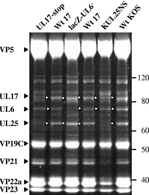
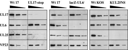
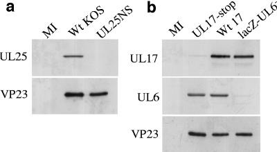
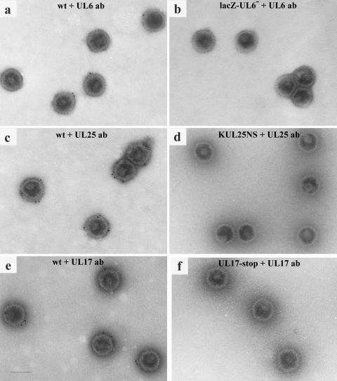
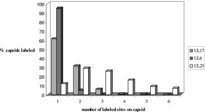
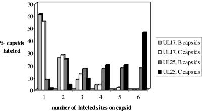
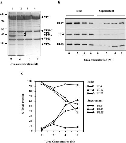
Herpes simplex virus type 1 packages its DNA genome into a precursor capsid, referred to as the procapsid. Of the three capsid-associated DNA-packaging proteins, UL17, UL25, and UL6, only UL17 and UL6 appear to be components of the procapsid, with UL25 being added subsequently. To determine whether the association of UL17 or UL25 with capsids was dependent on the other two packaging proteins, B capsids, which lack viral DNA but retain the cleaved internal scaffold, were purified from nonpermissive cells infected with UL17, UL25, or UL6 null mutants and compared with wild-type (wt) B capsids. In the absence of UL17, the levels of UL25 in the mutant capsids were much lower than those in wt B capsids. These results suggest that UL17 is required for efficient incorporation of UL25 into B capsids. B capsids lacking UL25 contained about twofold-less UL17 than wt capsids, raising the possibilities that UL25 is important for stabilizing UL17 in capsids and that the two proteins interact in the capsid. The distribution of UL17 and UL25 on B capsids was examined using immunogold labeling. Both proteins appeared to bind to multiple sites on the capsid. The properties of the UL17 and UL25 proteins are consistent with the idea that the two proteins are important in stabilizing capsid-DNA structures rather than having a direct role in DNA packaging.


Figures

FIG. 1.
FIG. 1.
Identification of packaging proteins UL6, UL17, and UL25 in wt virus and mutant B capsids. Purified B capsid proteins were separated by SDS-PAGE, and the proteins were detected by SYPRO orange staining. Capsid proteins are indicated on the left, and the positions of molecular weight markers (MagicMarkXP; in thousands) are shown on the right.
FIG. 2.
FIG. 2.
Analysis of UL6, UL17, and UL25 in wt HSV-1 strain 17, wt strain KOS, lacZ-UL6, KUL25NS, and UL17-stop capsids by Western blotting. Serial twofold dilutions of wt and mutant capsids that had been equalized on the basis of their VP23 content were prepared. The polypeptides were resolved by SDS-PAGE and blotted onto a nitrocellulose membrane. The blots were probed sequentially with antibodies UL6 MAb 175, UL17 MAb 203, UL25 MAb 166, and VP23 antibody Rab186 as indicated on the left side. In this experiment the UL25NS B capsid preparation was banded twice on a sucrose gradient to ensure minimal contamination with UL25NS empty capsids, lacking the scaffolding proteins.
FIG. 3.
FIG. 3.
Western blot analysis of proteins of MRUL17-stop and mutant ΔUL17 B capsids. For comparison, proteins of wt strain 17 and mutant UL17-stop B capsids were also included. Serial twofold dilutions of wt and mutant B capsids that had been equalized on the basis of their VP23 content were prepared. The polypeptides were resolved by SDS-PAGE and blotted onto a nitrocellulose membrane. The blots were probed sequentially with antibodies UL17 MAb 203, UL25 MAb 166, and VP23 polyclonal antibody 186 as indicated on the left side.
FIG. 4.
FIG. 4.
Specificity of UL6, UL17, and UL25 rabbit polyclonal antibodies. BHK cells were infected with HSV-1 wt strain 17, wt strain KOS, UL17-stop mutant, lacZ-UL6, or UL25NS or were mock infected (MI). At 24 h postinfection the cells were harvested and the proteins were separated by SDS-PAGE. Western blot analysis was carried out, probing the KOS, UL25NS, and mock-infected cell profiles sequentially with UL25 and VP23 polyclonal antibodies (a) and strain 17, lacZ-UL6, UL17-stop, and mock-infected cell profiles sequentially with UL17, UL6, and VP23 polyclonal antibodies (b).
FIG. 5.
FIG. 5.
Electron micrographs of wt and mutant capsids treated with UL6 antibody (a and b), UL25 antibody (c and d), or UL17 antibody (e and f). Capsids were incubated with the packaging protein antibody and subsequently treated with goat anti-rabbit IgG coupled to 10-nm colloidal gold. Capsids were negatively stained and examined under the electron microscope. Bar, 100 nm.
FIG. 6.
FIG. 6.
Distribution of UL25, UL17, and UL6 on wt B capsids. The percentage of labeled wt B capsids (treated with UL25, UL17, or UL6 antibodies) was plotted against the number of sites labeled with gold particles on each capsid.
FIG. 7.
FIG. 7.
Comparison of the distribution of UL25 and UL17 on wt B and C capsids. The percentage of labeled B or C wt strain 17 capsids (treated with UL25 or UL17 antibodies) was plotted against the number of sites labeled with gold particles on each capsid. The last column represents all capsids with six or more sites labeled with gold particles. The capsids examined were as follows: 164 wt B capsids treated with UL25 antibody were analyzed, 96% of which were labeled with gold; 137 wt B capsids treated with UL17 antibody were analyzed, 61% of which were labeled with gold; 147 wt C capsids treated with UL25 antibody were analyzed, 99% of which were labeled with gold; and 136 wt C capsids treated with UL17 were analyzed, 69% of which were labeled with gold.
FIG. 8.
FIG. 8.
Treatment of capsids with urea. Purified wt HSV-1 strain 17 B capsids were treated with various concentrations of urea for 1 h and concentrated by centrifugation. The proteins from the capsid pellets were resolved by SDS-PAGE and stained with Coomassie blue (a). Molecular masses in kilodaltons are shown at the left. In addition, the separated capsid proteins from the pellets and the supernatants were blotted onto nitrocellulose and the UL6, UL17, and UL25 packaging proteins were identified by Western blotting (b). The protein bands in the exposed film from the Western blot experiment shown in Fig. 7b were quantified by densitometric analysis of digital images (c).

References

    1. Addison, C., F. J. Rixon, and V. G. Preston. 1990. Herpes simplex virus type-1 UL28 gene product is important for the formation of mature capsids. J. Gen. Virol. 71:2377-2384. - PubMed
    1. Adelman, K., B. Salmon, and J. D. Baines. 2001. Herpes simplex virus DNA packaging sequences adopt novel structures that are specifically recognized by a component of the cleavage and packaging machinery. Proc. Natl. Acad. Sci. USA 98:3086-3091. - PMC - PubMed
    1. Beard, P. M., N. S. Taus, and J. D. Baines. 2002. DNA cleavage and packaging proteins encoded by genes UL28, UL15, and UL33 of herpes simplex virus type 1 form a complex in infected cells. J. Virol. 76:4785-4791. - PMC - PubMed
    1. Black, L. W. 1989. DNA packaging in dsDNA bacteriophages. Annu. Rev. Microbiol. 43:267-292. - PubMed
    1. Booy, F. P., W. W. Newcomb, B. L. Trus, J. C. Brown, T. S. Baker, and A. C. Steven. 1991. Liquid-crystalline, phage-like packing of encapsidated DNA in herpes simplex virus. Cell 64:1007-1015. - PMC - PubMed

Publication types

Substances

LinkOut - more resources