. 2006 Mar;80(5):2170-82.
doi: 10.1128/JVI.80.5.2170-2182.2006.

RNA secondary structure in the coding region of dengue virus type 2 directs translation start codon selection and is required for viral replication

Affiliations

RNA secondary structure in the coding region of dengue virus type 2 directs translation start codon selection and is required for viral replication

Karen Clyde et al. J Virol. 2006 Mar.

Abstract

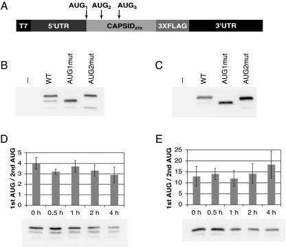
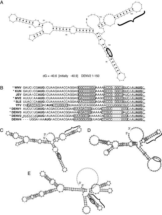
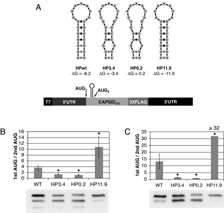
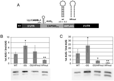
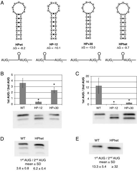
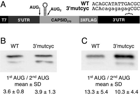
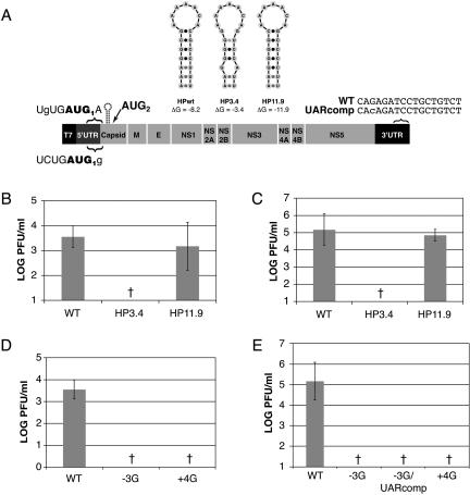
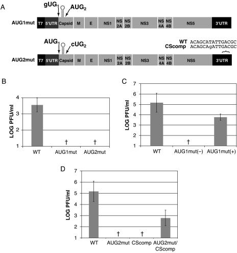
Dengue virus is a positive-strand RNA virus and a member of the genus Flavivirus, which includes West Nile, yellow fever, and tick-borne encephalitis viruses. Flavivirus genomes are translated as a single polyprotein that is subsequently cleaved into 10 proteins, the first of which is the viral capsid (C) protein. Dengue virus type 2 (DENV2) and other mosquito-borne flaviviruses initiate translation of C from a start codon in a suboptimal context and have multiple in-frame AUGs downstream. Here, we show that an RNA hairpin structure in the capsid coding region (cHP) directs translation start site selection in human and mosquito cells. The ability of the cHP to direct initiation from the first start codon is proportional to its thermodynamic stability, is position dependent, and is sequence independent, consistent with a mechanism in which the scanning initiation complex stalls momentarily over the first AUG as it begins to unwind the cHP. The cHP of tick-borne flaviviruses is not maintained in a position to influence start codon selection, which suggests that this coding region cis element may serve another function in the flavivirus life cycle. Here, we demonstrate that the DENV2 cHP and both the first and second AUGs of C are necessary for efficient viral replication in human and mosquito cells. While numerous regulatory elements have been identified in the untranslated regions of RNA viral genomes, we show that the cHP is a coding-region RNA element that directs start codon selection and is required for viral replication.


Figures

FIG. 1.
FIG. 1.
Translation of DENV2 capsid initiates from multiple AUGs. RNAs consisting of the DENV2 5′ UTR, the first 270 nt of the capsid gene, a 3XFLAG epitope, and the DENV2 3′ UTR (C-FLAG) were transfected into Hep3B and C6/36 cells. (A) Schematic diagram of C-FLAG RNA constructs. (B) Anti-FLAG immunoblot of C-FLAG-transfected Hep3B cells at 4 h posttransfection. (C) Anti-FLAG blot of transfected C6/36 cells at 24 h posttransfection. (D) Anti-FLAG immunoblot of Hep3B cells treated with 10 μg/ml cycloheximide (CHX) at 3 h posttransfection, incubated for 30 min, and lysed at the time indicated. The efficiency of codon usage is expressed as a ratio of the higher-molecular-weight C-FLAG isoform (1st AUG) to the lower-molecular-weight isoform (2nd AUG), and the mean ratio and SD were calculated from four experiments. (E) Anti-FLAG immunoblot of C6/36 cells treated as in panel D with the addition of CHX at 18 h posttransfection. The graph is as in panel D; the mean ratio and SD were calculated from five experiments. −, no RNA transfection (control).
FIG. 2.
FIG. 2.
A conserved hairpin element is predicted among mosquito- and tick-borne flaviviruses. (A) The RNA secondary structure of the first 150 nt of DENV2 was predicted by mfold; the start codon is circled, and the cHP is indicated by a bracket. (B) Alignment of start codons and the first in-frame AUG of mosquito-borne flaviruses, with predicted cHP stem regions outlined by rectangles. The −3 and +4 positions are indicated in gray letters. Viruses with a poor initiation context are indicated by asterisks. 5′ CS regions are indicated by solid arrows. The DENV2 5′ UAR region is indicated by a dashed arrow. (C) Phylogenetic consensus structure based on aligned sequences of DENV1, DENV2, DENV3, and DENV4 as computed by RNAalifold, with the start codon circled and the cHP indicated with a bracket. Covariant residues are circled. (D) Phylogenetic consensus structure of the mosquito-borne Japanese encephalitis serogroup viruses, WNV, Kunjin virus, JEV, St. Louis encephalitis virus, and Murray valley encephalitis virus, as in panel C. (E) Phylogenetic consensus structure of the tick-borne viruses: tick-borne encephalitis, Omsk hemorrhagic fever, Kyasanur Forest disease, and Powassan viruses, as in panel C.
FIG. 3.
FIG. 3.
The DENV2 cHP regulates translation initiation site selection. (A) Schematic diagram of constructs used to test different HP free energies. (B) Anti-FLAG immunoblot of C-FLAG-transfected Hep3B cells at 4 h posttransfection. The efficiency of codon usage is expressed as a ratio of the higher-molecular-weight C-FLAG isoform (1st AUG) to the lower-molecular-weight isoform (2nd AUG). The error bars indicate SDs; the data are derived from four experiments. (C) Anti-FLAG immunoblot of transfected C6/36 cells at 24 h posttransfection as in panel B. The graph is as in panel B; the data represent the averages of three experiments. The ratio of 32 reflects the maximum difference detectable by immunoblotting under the conditions described. *, P < 0.001 relative to wt.
FIG. 4.
FIG. 4.
Comparison of first start codon selection by DENV2 cHP to selection by improved start codon context. (A) Schematic diagram of constructs used to test the efficiency of first start codon selection by the cHP or by initiation context. (B) Anti-FLAG immunoblot of C-FLAG-transfected Hep3B cells at 4 h posttransfection. The efficiency of codon usage is expressed as a ratio of the higher-molecular-weight C-FLAG isoform (1st AUG) to the lower-molecular-weight isoform (2nd AUG). The error bars indicate SDs; the data are derived from five experiments. (C) Anti-FLAG immunoblot of transfected C6/36 cells at 24 h posttransfection as in panel B. The graph is as in panel B; the data represent the averages of four experiments. *, P < 0.05 relative to wt; **, P < 0.01 relative to −3G/HPmut.
FIG. 5.
FIG. 5.
The DENV2 cHP directs start codon selection via a position-dependent, sequence-independent mechanism. (A) Schematic diagrams of constructs used to test the position and sequence dependence of the DENV2 cHP in regulating start site selection. (B) Anti-FLAG immunoblot of C-FLAG-transfected Hep3B cells at 4 h posttransfection. The efficiency of codon usage is expressed as a ratio of the higher-molecular-weight C-FLAG isoform (1st AUG) to the lower-molecular-weight isoform (2nd AUG). The error bars indicate SDs; the data are derived from three experiments. (C) Anti-FLAG immunoblot of transfected C6/36 cells at 24 h posttransfection as in panel B. The graph is as in panel B; the data represent the averages of three experiments. (D) Anti-FLAG immunoblot of transfected Hep3B cells at 4 h posttransfection as in panel B. The mean ratio and SD are calculated from four experiments. (E) Anti-FLAG immunoblot of transfected C6/36 cells at 24 h posttransfection as in panel B. The table is as in panel D; the data represent the averages of four experiments. The ratio of 32 reflects the maximum difference detectable by immunoblotting under the conditions described. *, P < 0.01 relative to wt.
FIG. 6.
FIG. 6.
5′-3′ cyclization is not required for cHP-mediated start codon selection. (A) Schematic diagram of constructs used to test the impact of potential 5′-3′ cyclization on initiation site selection. (B) Anti-FLAG immunoblot of C-FLAG-transfected Hep3B cells at 4 h posttransfection. The efficiency of codon usage is expressed as a ratio of the higher-molecular-weight C-FLAG isoform (1st AUG) to the lower-molecular-weight isoform (2nd AUG). The data are derived from three experiments. (C) Anti-FLAG immunoblot of transfected C6/36 cells at 24 h posttransfection as in panel B. The table is as in panel B; the data represent the averages of three experiments.
FIG. 7.
FIG. 7.
The cHP is required for efficient DENV2 replication. In vitro-transcribed IC RNAs were transfected into Hep3B and C6/36 cell monolayers, and viral replication was assessed after 72 h by plaque assay. The titers were normalized to transfection efficiency as determined by qRT-PCR at 2 h posttransfection. (A) Schematics of IC variants utilized to study the role of the cHP and of the nucleotides that make up the DENV2 initiation context. (B) Viral titers are expressed as PFU per ml from IC-transfected Hep3B cells. One log unit reflects the limit of detection of a standard plaque assay. The error bars indicate SD; the data are derived from at least four experiments. (C) Viral titers from IC-transfected C6/36 cells. The graph is as in panel B; the data are derived from at least four experiments. (D) Viral titers from IC-transfected Hep3B cells. The graph is as in panel B; the data are derived from at least four experiments. (E) Viral titers from IC-transfected C6/36 cells. The graph is as in panel B; the data are derived from at least three experiments. †, not detectable.
FIG. 8.
FIG. 8.
Both the first and second AUGs of capsid play a role in the viral life cycle. In vitro-transcribed IC RNAs were transfected into Hep3B and C6/36 cell monolayers, and viral replication was assessed at 72 h by plaque assay. The titers were normalized to transfection efficiency as determined by qRT-PCR at 2 h posttransfection. (A) Schematic of IC variants utilized to study the roles of the first two AUGs in the capsid coding region. (B) Viral titers expressed as PFU/ml from IC-transfected Hep3B cells. One log unit reflects the limit of detection of a standard plaque assay. The error bars indicate SDs; the data are derived from at least three experiments. (C) Viral titers from IC-transfected C6/36 cells. The graph is as in panel B; the data are derived from at least five experiments. AUG1mut (−), not detectable in two of five experiments; AUG1mut (+), titerable virus detected in three of five experiments. (D) Viral titers from IC-transfected C6/36 cells. The graph is as in panel B; the data are derived from at least three experiments. †, not detectable.

References

    1. Alvarez, D. E., A. L. De Lella Ezcurra, S. Fucito, and A. V. Gamarnik. 2005. Role of RNA structures present at the 3′UTR of dengue virus on translation, RNA synthesis, and viral replication. Virology 339:200-212. - PubMed
    1. Alvarez, D. E., M. F. Lodeiro, S. J. Luduena, L. I. Pietrasanta, and A. V. Gamarnik. 2005. Long-range RNA-RNA interactions circularize the dengue virus genome. J. Virol. 79:6631-6643. - PMC - PubMed
    1. Bampi, C., S. Jacquenet, D. Lener, D. Decimo, and J. L. Darlix. 2004. The chaperoning and assistance roles of the HIV-1 nucleocapsid protein in proviral DNA synthesis and maintenance. Int. J. Biochem. Cell. Biol. 36:1668-1686. - PubMed
    1. Bell, J. R., R. M. Kinney, D. W. Trent, E. M. Lenches, L. Dalgarno, and J. H. Strauss. 1985. Amino-terminal amino acid sequences of structural proteins of three flaviviruses. Virology 143:224-229. - PubMed
    1. Bulich, R., and J. G. Aaskov. 1992. Nuclear localization of dengue 2 virus core protein detected with monoclonal antibodies. J. Gen. Virol. 73:2999-3003. - PubMed

Publication types

LinkOut - more resources