. 2006 Mar;80(5):2234-42.
doi: 10.1128/JVI.80.5.2234-2242.2006.

Transcriptional origin of Kaposi's sarcoma-associated herpesvirus microRNAs

Affiliations

Transcriptional origin of Kaposi's sarcoma-associated herpesvirus microRNAs

Xuezhong Cai et al. J Virol. 2006 Mar.

Abstract

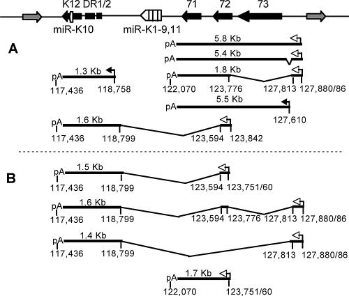
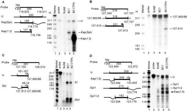
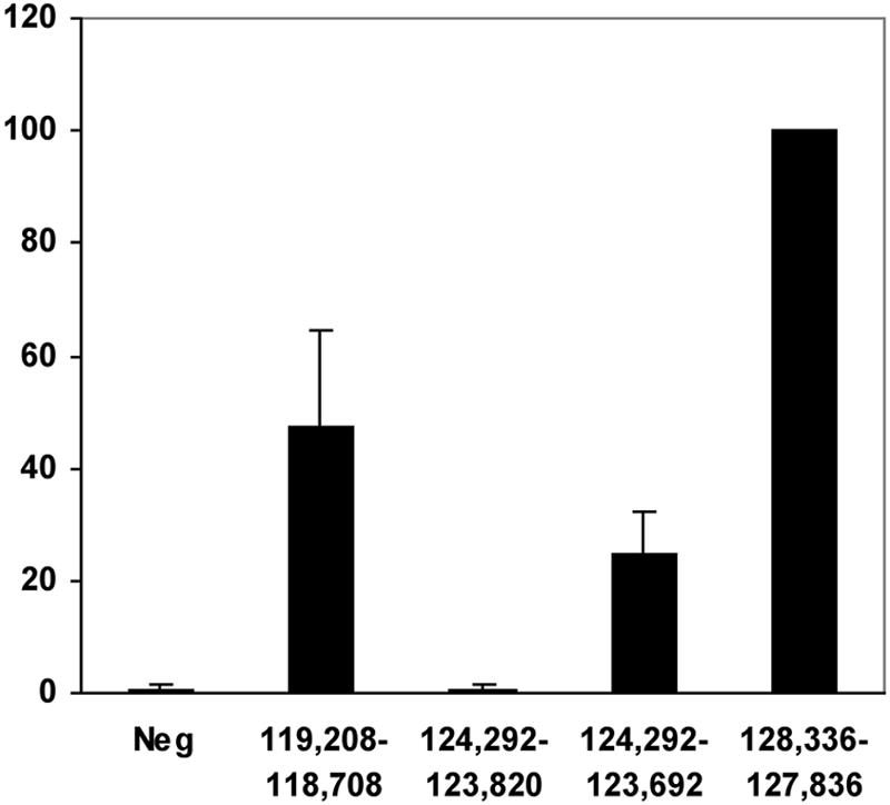
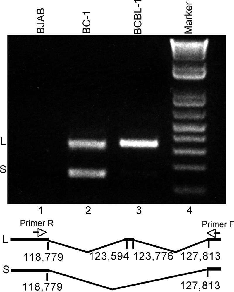
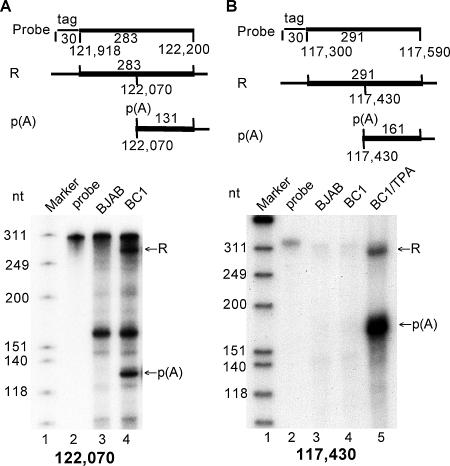
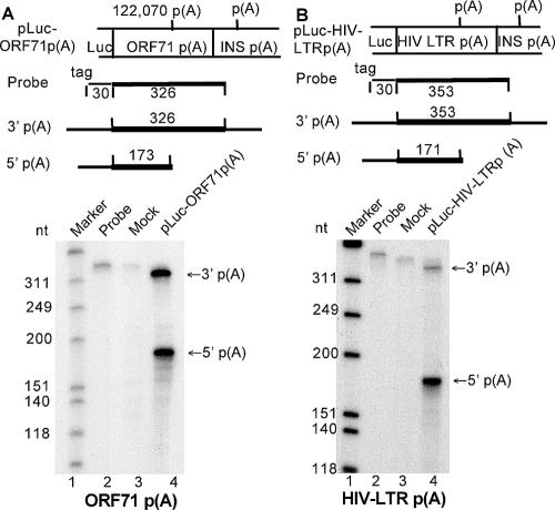
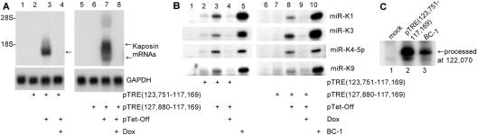
Kaposi's sarcoma-associated herpesvirus (KSHV) encodes 11 distinct microRNAs, all of which are found clustered within the major latency-associated region of the KSHV genome in the same transcriptional orientation. Because the KSHV microRNAs are all expressed in latently infected cells and are largely unaffected by induction of lytic replication, it appeared probable that they would be processed out of KSHV transcripts that are derived from a latent promoter(s) present in this region. Here, we define three latent transcripts, derived from two distinct KSHV latent promoters, that function as both KSHV primary microRNA precursors and as kaposin pre-mRNAs. These activities require the readthrough of a leaky viral polyadenylation signal located at nucleotide 122070 in the KSHV genome. In contrast, recognition of this polyadenylation signal gives rise to previously identified mRNAs that encode the KSHV open reading frames (ORFs) 71, 72 and 73 proteins as well as a novel unspliced KSHV mRNA that encodes only ORF72 and ORF71. Thus, transcripts initiating at the two latent promoters present in the KSHV latency-associated region can undergo two entirely distinct fates, i.e., processing to give a kaposin mRNA and viral microRNAs on the one hand or expression as KSHV ORF71, ORF72, or ORF73 mRNAs on the other, depending on whether the viral polyadenylation site located at position 122070 is ignored or recognized, respectively.


Figures

FIG. 1.
FIG. 1.
Transcription of the latency-associated region of the KSHV genome. The KSHV latency-associated region encodes four proteins, indicated by black boxes, as well as 11 miRNAs (white boxes) and is flanked by lytic genes (gray boxes). In panel A, previously reported promoters, splice sites, and poly(A) addition sites are indicated. Lytic promoters are indicated by black arrows and latent promoters by white arrows. Novel KSHV transcripts identified in this report are shown in panel B. Sequence coordinates are derived from the KSHV genome sequence obtained from BC-1 cells (GenBank accession number NC_003409).
FIG. 2.
FIG. 2.
Characterization of promoters located in the KSHV latency-associated region. (A) This RPA utilized a probe designed to detect RNAs that initiate transcription at position 118758 as well as spliced (Spl) and unspliced (U) KSHV RNAs that initiate 5′ to position 118911. The RNA samples used were from the uninfected B-cell line BJAB (lane 3) or from the latently KSHV-infected cell line BC-1. BC-1 cells were cultured under conditions that allow only ∼0.9% of cells to spontaneously enter lytic KSHV replication (lane 4) or were treated with TPA, which induces lytic KSHV replication in ∼20% of the BC-1 cells (lane 5). (B) Similar to panel A, except that this 348-nucleotide RPA probe was designed to detect transcripts initiating at the 127610 promoter. Transcripts initiating at 127610 would rescue a 221-nucleotide probe fragment, while RNAs initiating at the 127880/86 promoter would rescue a 318-nucleotide probe fragment. (C) Similar to panel A, except that the RPA probe used detects transcripts that initiate at the 127880/86 promoter. Unspliced RNAs (U) and RNAs that utilize the 5′ splice site at 127813 (Spl) were detected. (D) Similar to panel A except that this RPA probe was designed to detect transcripts adjacent to the proposed 123842 cap site. Transcripts initiating at the 127880/86 promoter should give rise to 309- and 213-nucleotide rescued fragments representing unspliced (U) and singly spliced (Spl) RNAs, respectively. A 183-nucleotide probe fragment is predicted if a transcript initiating at 127880/86 is doubly spliced (Spl1+2). No band at the predicted size of 249 nucleotides, reflecting transcription initiation at 123842, was detected. The provenance of the ∼167- and ∼158-nucleotide probe fragments is discussed in the text.
FIG. 3.
FIG. 3.
Biological activity of candidate KSHV promoter elements. Plasmids containing the indicated KSHV DNA fragments linked to the luciferase indicator gene were transfected into 293T cells and induced luciferase activities determined. A Renilla luciferase expression vector was cotransfected as an internal control. The data presented were normalized to the internal control and are given relative to the pTRE(128336-127836)Luc indicator plasmid, which was set at 100. The negative (Neg) control contains the antisense version of the 128336/127836 KSHV sequence linked to the luciferase gene. The average for three experiments with standard deviations is indicated.
FIG. 4.
FIG. 4.
Reverse transcription-PCR analysis of spliced kaposin mRNAs derived from the 127880/86 promoter. Total RNA derived from BJAB, BC-1, or BCBL-1 cells was subjected to reverse transcription using oligo(dT) primers and then PCR using primers extending from 118098 to 118718 (R) and from 127861 to 127879 (F) in the KSHV genome. This analysis yielded a large (L) fragment of 332 bp and a short (S) fragment of 149 bp. DNA sequencing demonstrated that these two fragments were derived from KSHV RNAs that had undergone alternative splicing as indicated.
FIG. 5.
FIG. 5.
Relative polyadenylation efficiency at the KSHV 117430 and 122070 polyadenylation sites in BC-1 cells. This RPA analysis uses probes that detect KSHV transcripts that read through (R) the 117430 or 122070 polyadenylation site or are processed [p(A)] at these sites. The RNA samples used were from control BJAB cells or from latently KSHV-infected BC-1 cells. The probe design and the predicted rescued probe fragments are indicated at the top.
FIG. 6.
FIG. 6.
Analysis of processing efficiency at the KSHV 122070 poly(A) addition site. This analysis used indicator constructs in which ∼300-bp DNA fragments that include the entire KSHV 122070 polyadenylation site (A) or the human immunodeficiency virus type 1 long terminal repeat (HIV-1 LTR) polyadenylation site [p(A)] (B) were introduced between a luciferase indicator gene and a highly active genomic rat preproinsulin II (INS) polyadenylation site. The probes utilized distinguish between RNAs processed at the introduced 5′ polyadenylation site and those processed at the 3′ insulin polyadenylation site or that remain unprocessed.
FIG. 7.
FIG. 7.
Identification of KSHV pre-mRNAs that also function as primary miRNAs. Expression constructs consisting of a tetracycline-regulated promoter linked to either segments 123751 to 117169 or 127880 to 117169 of the KSHV genome were introduced into 293T cells together with the pTet-Off expression plasmid in the presence or absence of doxycycline. At 48 hours after transfection, cells were lysed and total RNA was isolated. (A) Northern analysis using a K12-specific probe. Endogenous glyceraldehyde-3-phosphate dehydrogenase (GAPDH) mRNA was used as a loading control. The introduced plasmids are indicated, as is whether doxycycline was added. Lanes 1 and 5 represent mock-transfected 293T cultures. (B) Primer extension assay performed using the same RNAs analyzed in panel A. The primers used are designed to detect the indicated KSHV miRNAs. BC-1 RNA (lanes 5 and 10) was used as a positive control. (C) RPA analysis of RNA derived from 293T cells transfected with pTRE(123751-117169) and pTet-Off (lane 2) or using BC-1 RNA (lane 3) and the RPA probe described in Fig. 5B. The indicated band represents RNA processed at the 122070 polyadenylation site.

References

    1. Bartel, D. P. 2004. MicroRNAs: genomics, biogenesis, mechanism, and function. Cell 116:281-297. - PubMed
    1. Brown, P. H., L. S. Tiley, and B. R. Cullen. 1991. Efficient polyadenylation within the human immunodeficiency virus type 1 long terminal repeat requires flanking U3-specific sequences. J. Virol. 65:3340-3343. - PMC - PubMed
    1. Cai, X., C. H. Hagedorn, and B. R. Cullen. 2004. Human miRNAs are processed from capped, polyadenylated transcripts that can also function as mRNAs. RNA 10:1957-1966. - PMC - PubMed
    1. Cai, X., S. Lu, Z. Zhang, C. M. Gonzalez, B. Damania, and B. R. Cullen. 2005. Kaposi's sarcoma-associated herpesvirus expresses an array of viral miRNAs in latently infected cells. Proc. Natl. Acad. Sci. USA 102:5570-5575. - PMC - PubMed
    1. Chendrimada, T. P., R. I. Gregory, E. Kumaraswamy, J. Norman, N. Cooch, K. Nishikura, and R. Shiekhattar. 2005. TRBP recruits the Dicer complex to Ago2 for miRNA processing and gene silencing. Nature 436:740-744. - PMC - PubMed

Publication types

LinkOut - more resources