. 2006 Mar;80(5):2267-79.
doi: 10.1128/JVI.80.5.2267-2279.2006.

A single intranasal inoculation with a paramyxovirus-vectored vaccine protects guinea pigs against a lethal-dose Ebola virus challenge

Affiliations

A single intranasal inoculation with a paramyxovirus-vectored vaccine protects guinea pigs against a lethal-dose Ebola virus challenge

Alexander Bukreyev et al. J Virol. 2006 Mar.

Abstract

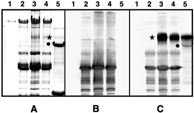
To determine whether intranasal inoculation with a paramyxovirus-vectored vaccine can induce protective immunity against Ebola virus (EV), recombinant human parainfluenza virus type 3 (HPIV3) was modified to express either the EV structural glycoprotein (GP) by itself (HPIV3/EboGP) or together with the EV nucleoprotein (NP) (HPIV3/EboGP-NP). Expression of EV GP by these recombinant viruses resulted in its efficient incorporation into virus particles and increased cytopathic effect in Vero cells. HPIV3/EboGP was 100-fold more efficiently neutralized by antibodies to EV than by antibodies to HPIV3. Guinea pigs infected with a single intranasal inoculation of 10(5.3) PFU of HPIV3/EboGP or HPIV3/EboGP-NP showed no apparent signs of disease yet developed a strong humoral response specific to the EV proteins. When these animals were challenged with an intraperitoneal injection of 10(3) PFU of EV, there were no outward signs of disease, no viremia or detectable EV antigen in the blood, and no evidence of infection in the spleen, liver, and lungs. In contrast, all of the control animals died or developed severe EV disease following challenge. The highly effective immunity achieved with a single vaccine dose suggests that intranasal immunization with live vectored vaccines based on recombinant respiratory viruses may be an advantageous approach to inducing protective responses against severe systemic infections, such as those caused by hemorrhagic fever agents.


Figures

FIG. 1.
FIG. 1.
Genome structures of recombinant HPIV3s expressing EV GP and NP. (A) Schematic diagrams of the genome of the HPIV3 vector, with insertion sites indicated by arrows (top) and HPIV3 vectors containing a transcription cassette encoding EV GP (middle) or two separate cassettes encoding GP and NP (bottom). The genomic length of each construct (in nucleotides) is indicated to the right. (B) Sequences flanking the EV GP (top) and NP (bottom) ORFs in the HPIV3 genome, shown in antigenomic or positive sense. The boundaries of the GP and NP inserts are indicated. HPIV3-specific gene start and gene end transcriptional signals are boxed, the three-nucleotide-long intergenic sequence is designated IG, and the initiation and stop codons of the GP and NP ORFs are in boldface, with the remainder of each ORF indicated by three dots. Restriction endonuclease sites used for cloning are indicated below their underlined sequences.
FIG. 2.
FIG. 2.
Adventitious mutations detected in the editing site of the EV GP ORF in recovered HPIV3/EboGP and HPIV3/EboGP-NP viruses. The top line shows the positive-sense sequence of part of the editing site in the unedited version of EV GP mRNA, with encoded amino acids (a.a.) and their sequence positions in GP indicated and the homo-oligomeric A tract underlined. The next line shows the edited version of the mRNA containing a tract of eight A residues; this is the version that initially was inserted into the HPIV3 vector but proved to be unstable. The next four lines illustrate sequences from recovered viruses containing a nucleotide substitution or a nucleotide deletion that disrupts this run of eight A residues. The sequence at the bottom is an engineered mutant version of the edited mRNA that contains two A→G substitutions that interrupt the run of eight A residues without affecting amino acid coding; this version was used to make the second set of HPIV3/EboGP and HPIV3/EboGP-NP viruses that were used in this study.
FIG. 3.
FIG. 3.
Western blot analysis of cell-associated EV GP and NP expressed by the recombinant vectors. LLC-MK2 cells were infected with the recombinant viruses, and total cell lysates were prepared 24 h later, separated by electrophoresis on 4 to 12% bis-Tris acrylamide gradient gels under denaturing and reducing conditions, and subjected to Western blot analysis. Replicate blots were analyzed with rabbit anti-EV antibodies (A), guinea pig anti-NP antibodies (B), and a mouse monoclonal antibody against actin as a loading control (C). Lanes: 1, molecular weight markers; 2, HPIV3; 3, HPIV3/EboGP; 4, HPIV3/EboGP-NP; 5, mock infection. Molecular masses of the marker proteins (in kilodaltons) are shown in the left margins, and the positions of EV GP1 (A) and NP (B) are indicated with arrows.
FIG. 4.
FIG. 4.
Incorporation of EV GP1 and GP2 into recombinant virus particles. Purified HPIV3 and HPIV3/EboGP were prepared by sedimentation on discontinuous sucrose gradients, separated by electrophoresis on 4 to 12% (top panel A and panel B) or on 10% (bottom panel A) bis-Tris acrylamide gradient gels under denaturing and reducing conditions, and subjected to Western blot analysis with guinea pig anti-GP antibodies (top panel A) or rabbit hyperimmune serum that had been raised against inactivated purified EV virions (bottom panel A). A second gel containing the same array of samples was analyzed by silver straining (B). Lanes: 1, molecular weight markers; 2, HPIV3; 3, HPIV3/EboGP. Molecular masses of the marker proteins (in kilodaltons) are shown in the left margins, and positions of EV GP1, GP2, and the major HPIV3 proteins are shown in the right margins.
FIG. 5.
FIG. 5.
Electron microscopy of virus particles. Shown are negative-stained images of HPIV3 (A), HPIV3/Ebo-GP (B), HPIV3/EboGP-NP (C), and EV (D). Glycoprotein peplomers are seen as a fringe along the outer edge of all virus particles, and extruding nucleocapsid is evident in panel B. Representative images are shown; in general, consistent differences between HPIV3 and its derivatives with EV gene inserts were not evident.
FIG. 6.
FIG. 6.
CPE produced by the indicated recombinant viruses on day 3 after infection in LLC-MK2 and Vero cell cultures. Infections were performed at an input MOI of 0.2 PFU per cell. Infection with HPIV3/EboGP-NP produced CPE indistinguishable from that with HPIV3/EboGP and therefore is not shown.
FIG. 7.
FIG. 7.
Multistep growth kinetics of the indicated recombinant viruses in LLC-MK2 and Vero cells. Cell monolayers were infected with the indicated viruses at an input MOI of 0.001 PFU per cell. Supernatants were harvested at 24-h intervals and flash frozen, and virus titers were determined in LLC-MK2 cells and expressed as PFU per milliliter. Mean values and standard error from triplicate samples are shown. These data are from a representative experiment out of a total of two independent experiments.
FIG. 8.
FIG. 8.
Mean body weights of the guinea pigs in the experimental groups that received the indicated recombinant viruses following the initial immunizing infection (A) and following the EV challenge on day 28 (B). The mean weights are plotted as percentages relative to the mean weights on the day of immunization (A) or challenge (B). Of the five animals in the control (HPIV3-vaccinated) group that were not sacrificed on day 7 postchallenge, two animals died on day 8, one animal on day 9, and two animals on day 10.
FIG. 9.
FIG. 9.
Specificity of guinea pig serum antibodies following the initial immunization. The following samples were separated in three replicate 7.5% SDS-polyacrylamide gels: mock-purified virus from uninfected cells (lane 1), purified HPIV3 (lane 2), purified HPIV3/EboGP (lane 3), purified HPIV3/GP-NP (lane 4), and purified EV (lane 5). One replicate gel was stained with Coomassie blue (A), and the other two were subjected to Western blot analysis with antiserum from a representative individual guinea pig taken on day 28 after immunization with HPIV3 (B) or HPIV3/EboGP-NP (C). The positions of EV GP1 and NP are shown by stars and circles, respectively, in panels A and C. The poor detection of GP1 in lane 5 of panel A probably reflects inefficient reactivity of the glycoprotein with the Coomassie stain due to the high density of the carbohydrate side chains. The lack of NP in lane 4 of panel C indicates that EV NP is not incorporated into the HPIV3 vector particles.
FIG. 10.
FIG. 10.
EV antigen detection in internal organs of guinea pigs on day 7 after the challenge. Shown are representative examples of IHC staining of spleen (A and D), liver (B and E), and lung (C and F) tissues from a control animal immunized with HPIV3 (A, B and C) and from an animal immunized with HPIV3/EboGP-NP (D, E and F). Areas of the section containing EV antigen are stained red. Bar, 100 μm.

References

    1. Baize, S., E. M. Leroy, M. C. Georges-Courbot, M. Capron, J. Lansoud-Soukate, P. Debre, S. P. Fisher-Hoch, J. B. McCormick, and A. J. Georges. 1999. Defective humoral responses and extensive intravascular apoptosis are associated with fatal outcome in Ebola virus-infected patients. Nat. Med. 5:423-426. - PubMed
    1. Baize, S., E. M. Leroy, E. Mavoungou, and S. P. Fisher-Hoch. 2000. Apoptosis in fatal Ebola infection. Does the virus toll the bell for immune system? Apoptosis 5:5-7. - PubMed
    1. Basler, C. F., X. Wang, E. Muhlberger, V. Volchkov, J. Paragas, H. D. Klenk, A. Garcia-Sastre, and P. Palese. 2000. The Ebola virus VP35 protein functions as a type I IFN antagonist. Proc. Natl. Acad. Sci. USA 97:12289-12294. - PMC - PubMed
    1. Bermingham, A., and P. L. Collins. 1999. The M2-2 protein of human respiratory syncytial virus is a regulatory factor involved in the balance between RNA replication and transcription. Proc. Natl. Acad. Sci. USA 96:11259-11264. - PMC - PubMed
    1. Bosio, C. M., M. J. Aman, C. Grogan, R. Hogan, G. Ruthel, D. Negley, M. Mohamadzadeh, S. Bavari, and A. Schmaljohn. 2003. Ebola and Marburg viruses replicate in monocyte-derived dendritic cells without inducing the production of cytokines and full maturation. J. Infect. Dis. 188:1630-1638. - PubMed

Publication types

MeSH terms