. 2006 Mar;80(5):2309-17.
doi: 10.1128/JVI.80.5.2309-2317.2006.

Structural characterization of the UL25 DNA-packaging protein from herpes simplex virus type 1

Affiliations

Structural characterization of the UL25 DNA-packaging protein from herpes simplex virus type 1

Brian R Bowman et al. J Virol. 2006 Mar.

Abstract

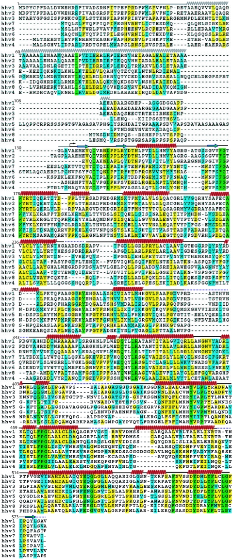
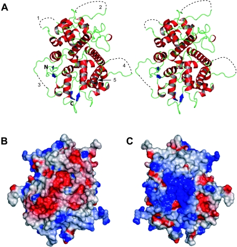
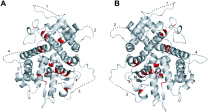
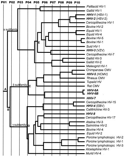
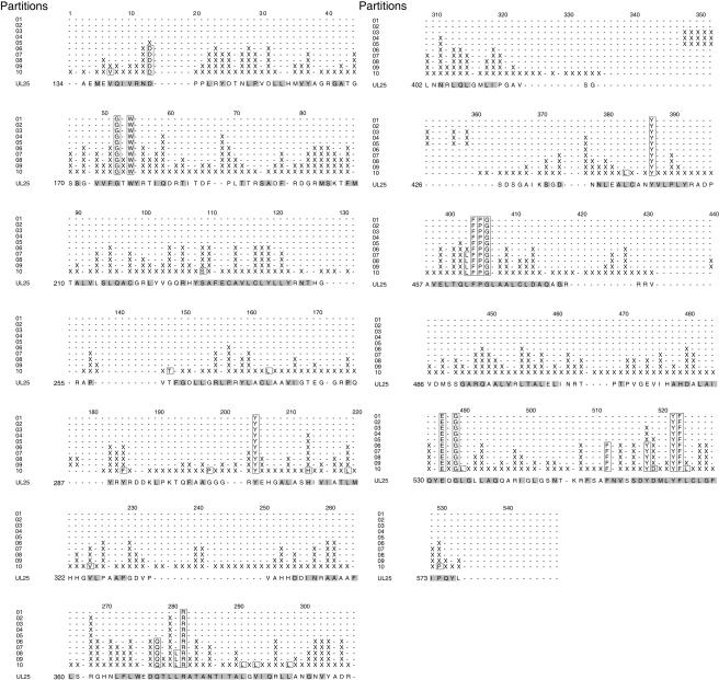
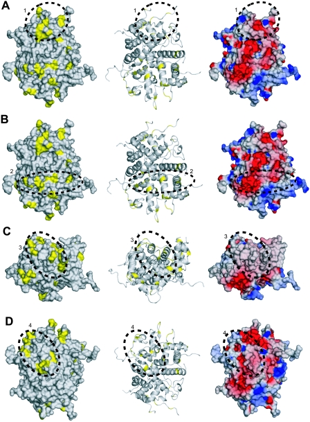
Herpesviruses replicate their double stranded DNA genomes as high-molecular-weight concatemers which are subsequently cleaved into unit-length genomes by a complex mechanism that is tightly coupled to DNA insertion into a preformed capsid structure, the procapsid. The herpes simplex virus type 1 UL25 protein is incorporated into the capsid during DNA packaging, and previous studies of a null mutant have demonstrated that its function is essential at the late stages of the head-filling process, either to allow packaging to proceed to completion or for retention of the viral genome within the capsid. We have expressed and purified an N-terminally truncated form of the 580-residue UL25 protein and have determined the crystallographic structure of the region corresponding to amino acids 134 to 580 at 2.1-Angstroms resolution. This structure, the first for any herpesvirus protein involved in processing and packaging of viral DNA, reveals a novel fold, a distinctive electrostatic distribution, and a unique "flexible" architecture in which numerous flexible loops emanate from a stable core. Evolutionary trace analysis of UL25 and its homologues in other herpesviruses was used to locate potentially important amino acids on the surface of the protein, leading to the identification of four putative docking regions for protein partners.


Figures

FIG. 1.
FIG. 1.
Sequence alignment of UL25 homologues from the eight human herpesviruses. The residues are colored green (completely conserved), yellow (identical residues), and cyan (similar residues). The secondary-structure elements are indicated above the sequence in red (helix), blue (strand), black (loop), and dotted line (disordered). The arrow indicates the position of the first ordered N-terminal residue (A134), and the helix and loop regions predicted for residues 1 to 133 are shown as gray wavy and straight lines, respectively.
FIG. 2.
FIG. 2.
The UL25nt structure. (A) Stereo view of the ribbon representation of the backbone structure. The secondary-structure elements are colored red (α-helices), blue (β-strands), and green (loops). The dotted black lines (1 to 4) represent the disordered loops, labeled for orientation reference. N and C labels identify the amino- and carboxy-terminal ends, respectively. (B) Electrostatic surface representation of the electronegative face of UL25nt. (C) A 180° rotation of the view in panel B, showing the electropositive face of UL25nt.
FIG. 3.
FIG. 3.
Conserved residues of UL25nt. (A) Ribbon diagram of UL25nt, with the conserved residues in Fig. 1 in red. (B) A 180° rotation of the view in panel A.
FIG. 4.
FIG. 4.
Evolutionary trace of herpesvirus UL25 homologues. P01 to P10 represent the different partitions of the ET phylogenetic tree. The branches defining the three herpesvirus subfamilies are labeled α, β, and γ. Key nodes discussed in the text are identified by A to D. Virus species names in boldface represent human viruses.
FIG. 5.
FIG. 5.
Evolutionary trace residues for partitions 01 to 10. The trace residues belonging to a given partition occur in the horizontal row corresponding to the partition. The conserved residues are outlined in a box, the class-specific residues are demarcated by an X, and the dashes reflect gaps in the alignment.
FIG. 6.
FIG. 6.
Evolutionary trace analysis. (A) The left and middle panels are a space-filling model and a ribbon trace, respectively, of UL25nt, with the class-specific exterior residues identified by the evolutionary trace shown in yellow. Cluster 1 is circled and identified with a 1. This cluster is also depicted in the electrostatic surface potential representation of the UL25nt structure (right). All three views are the same. (B, C, and D) Corresponding depictions of clusters 2, 3, and 4, respectively.

References

    1. Addison, C., F. J. Rixon, J. W. Palfreyman, M. O'Hara, and V. G. Preston. 1984. Characterisation of a herpes simplex virus type 1 mutant which has a temperature-sensitive defect in penetration of cells and assembly of capsids. Virology 138:246-259. - PubMed
    1. Adelman, K., B. Salmon, and J. D. Baines. 2001. Herpes simplex virus DNA packaging sequences adopt novel structures that are specifically recognized by a component of the cleavage and packaging machinery. Proc. Natl. Acad. Sci. USA 98:3086-3091. - PMC - PubMed
    1. Ali, M. A., B. Forghani, and E. M. Cantin. 1996. Characterization of an essential HSV-1 protein encoded by the UL25 gene reported to be involved in virus penetration and capsid assembly. Virology 216:278-283. - PubMed
    1. Bailey, S. 1994. The CCP4 suite—programs for protein crystallography. Acta Crystallogr. D 50:760-763. - PubMed
    1. Beard, P. M., N. S. Taus, and J. D. Baines. 2002. DNA cleavage and packaging proteins encoded by genes U(L)28, U(L)15, and U(L)33 of herpes simplex virus type 1 form a complex in infected cells. J. Virol. 76:4785-4791. - PMC - PubMed

Publication types

Associated data

LinkOut - more resources