Review
. 2006 Apr;147(8):839-53.
doi: 10.1038/sj.bjp.0706689.

Pharmacological insights obtained from structure-function studies of ionotropic glutamate receptors

Affiliations
Review

Pharmacological insights obtained from structure-function studies of ionotropic glutamate receptors

Philip E Chen et al. Br J Pharmacol. 2006 Apr.

Abstract

Ionotropic glutamate receptors mediate the vast majority of fast excitatory synaptic transmission in the CNS. Elucidating the structure of these proteins is central to understanding their overall function and in the last few years a tremendous amount of knowledge has been gained from the crystal structures of the ligand-binding domains of the receptor protein. These efforts have enabled us to unravel the possible mechanisms of partial agonism, agonist selectivity and desensitization. This review summarizes recent data obtained from structural studies of the binding pockets of the GluR2, GluR5/6, NR1 and NR2A subunits and discusses these studies together with homology modelling and molecular dynamics simulations that have suggested possible binding modes for full and partial agonists as well as antagonists within the binding pocket of various ionotropic glutamate receptor subunits. Comparison of the ligand-binding pockets suggests that the ligand-binding mechanisms may be conserved throughout the glutamate receptor family, although agonist selectivity may be explained by a number of features inherent to the AMPA, kainate and NMDA receptor-binding pockets such as steric occlusion, cavity size and the contribution of water-bridged interactions.


Figures
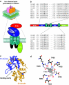
Figure 1
Figure 1
(a) Proposed iGluR structure and topology. iGluRs are thought to exhibit a tetrameric stoichiometry in a ‘dimer of dimers' configuration. NMDARs consist of two NR1 and NR2 subunits (though some may contain NR3 subunits), while AMPARs and kainate receptors can exist in either homomeric or heteromeric configurations. The proposed topological structure of an iGluR subunit is shown in greater detail in the cartoon below. The subunit is composed of a number of functional domains: an extracellular amino terminal domain (ATD), a ligand-binding region (S1 and S2), three transmembrane domains (M1, 3 and 4) and a re-entrant loop (M2) and a carboxyl terminal domain (CTD). (b) Partial amino-acid alignments highlighting the location of known ligand-binding residues (bold) identified by the crystal structures of GluR2, GluR5, GluR6 and NR1 S1S2 domains. Analogous residues in the NR2A and NR2B subunits are included for comparison. The partial sequence alignments for the NR3A and 3B subunits are also included; in both subunits, both essential threonine residues in the S1- and S2-binding domains are absent. Numbering is according to the mature polypeptide (Monyer et al., 1992). For GluR5 and GluR6 numbering is according to Mayer (2005a). (c) Structure of the S1S2 ligand-binding pocket for GluR2 in complex with kainate. The S1 (blue)- and S2 (gold)-binding domains form a hinged clamshell-like structure with the ligand-binding cavity nested between both regions. GluR2 coordinates were obtained from Armstrong et al., 1998 (PDB accession code-1gr2) and visualized using RasMol software. (d) A schematic representation of the contact residues within the GluR2 S1S2-binding pocket in direct contact with glutamate (open circles). Hydrogen bonds formed between binding pocket side chains and glutamate are shown as dashed lines. The residues Tyr450 and Glu705 appear ‘above' and ‘below' the complexed ligand within the binding site as if one was looking into the pocket from the top. In the GluR6 binding scheme, the Thr480 and Ser654 residues are replaced by two alanines (Ala 487 and Ala 658) reducing the stability of the GluR6–glutamate complex. Carbon atoms are black, oxygen, red and nitrogen, blue. Key for standard abbreviations of amino acids: A, Ala; D, Asp; E, Glu; F, Phe; G, Gly; H, His; I, Iso; K, Lys; L, Leu; M, Met; N, Asn; P, Pro; Q, Gln; R, Arg; S, Ser; T, Thr; V, Val; W, Trp and Y, Tyr. Panels (c) and (d) reprinted with permission from Erreger et al. (2004) (Critical Reviews in Neurobiology 16, 187–224. © 2004 Begell House Inc.).
Figure 2
Figure 2
Chemical structures of the various agonists and antagonists that interact with the glutamate-binding site, in NMDA, AMPA and kainate receptor subunits and the glycine-binding site of the NR1 NMDA receptor subunit.
Figure 3
Figure 3
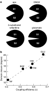
(a) Cartoon to illustrate the fact that different ligands cause difference amounts of domain closure of GluR2 receptor subunits. In the unliganded (apo) state the separation between the S1 and S2 domains is at its greatest. Antagonists such as DNQX cause only small changes in the degree of domain closure. Partial agonists such as the 5-substituted willardiine cause greater domain closure than is seen with antagonists but less than that seen with full agonists such as glutamate. (b) The extent of domain closure is correlated to the efficiency with which agonists open AMPARs containing the GluR2 receptor subunit. Key to abbreviations: GLU=glutamate; HW=willardiine; FW=fluorowillardiine; BrW=bromowillardiine; IW=iodowillardiine. Panel (b) adapted and reprinted, with permission, from Jin et al. (2003) (Nature Neuroscience 6, 803–810. © 2003 Nature Publishing Group (www.nature.com/)).
Figure 4
Figure 4
(a) Superimposition of structures showing glutamate docked in the ligand-binding site of NR2A NMDA receptor subunits (grey) and GluR2 AMPA receptor subunits (pink). (b) Illustration showing the residues directly and indirectly interacting with glutamate in the crystal structure of the NR2AS1S2-binding pocket. Dashed lines represent hydrogen bonds or salt bridges and the water-mediated network is formed by two water molecules (W1 and W2). In contrast from previous NR2 homology models, the Asp731 residue does not directly interact with the α-amino group of glutamate, while the Tyr730 residue interacts directly with the γ-carboxyl group of glutamate. Reprinted with permission from Furukawa et al. (2005) (Nature 438, 185–192. © 2005 Nature Publishing Group (www.nature.com/)).
Figure 5
Figure 5
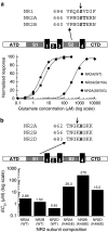
(a) Different mutations at one of the serine residues known to be involved in glutamate binding can exert drastically different effects on glutamate potency (location shown by arrowhead in the alignment above). Glutamate concentration–response curves for NR2A wild type, NR2A(S670A)- and NR2A(S670G)-containing receptors. The NR2A(S670A) mutation only increased glutamate EC50 to 4.97 μM (approximately two-fold more than wild type); however, the NR2A(S670G) mutation reduced glutamate potency by 145-fold to 421 μM. This compares well with observations of the equivalent mutations in NR2B-containing receptors (Laube et al., 1997; 2004). Data from Anson et al. (1998) and Chen et al. (2005). Error bars omitted for clarity. (b) The equivalent lysine to glutamic acid mutation within the S1 domain can produce differential glutamate sensitivities among NR2A-, NR2B- and NR2D-containing receptors. The bar graph compares mean glutamate EC50 values for wild-type NR2A, NR2B- or NR2D-containing receptors and their Lys to Glu mutant counterparts, NR2A(K465E), NR2B(K459E) and NR2D(K486E). The NR2B(K459E) mutation reduces glutamate potency by about 180-fold compared to NR2A(K465E) and NR2D(K486E) (10- and 34-fold). Location of the lysine residue (arrowhead) among the NR2 subunits is highlighted above. NR2A and NR2B data from Laube et al. (1997) and Anson et al. (1998) respectively, NR2D data from Chen, 2000.
Figure 6
Figure 6
(a) Possible mechanism underlying NMDA selectivity as judged by comparing both GluR2 and NR2 pockets. The position of the side chain from Glu705 in GluR2 would sterically occlude the N-methyl group of NMDA (yellow sphere), whereas in NR2A the side chain group of the Asp731 residue would not hinder NMDA binding. (b) Superimposing the ball and stick schemes of the homology model of NR2A-NMDA from Chen et al. (2005) with the GluR2S1S2-glutamate crystal structure reveals that a methionine residue (Met 708) in GluR2 may sterically hinder the N-methyl group of NMDA. In NR2A, this residue is replaced by the smaller valine residue facilitating NMDA binding. The GluR2 backbone is shown in grey, while the NR2A S1 and S2 regions are coloured orange and green, respectively. Equivalent GluR2 and NR2A residues are labelled in white or green text, respectively. (c) Surface profiles of the GluR2 (light brown) and NR2A schemes (dark brown) shown in (b). Location of Met708 in GluR2 is shown by the white arrow. Panel (a) reprinted with permission from Furukawa et al. (2005) (Nature 438, 185–192. © 2005 Nature Publishing Group (www.nature.com/)). Panels (b) and (c) reprinted, with permission, from Chen et al. (2005) (Molecular Pharmacology 67, 1470–1484. © 2005 American Society for Pharmacology and Experimental Therapeutics).
Figure 7
Figure 7
(a) Examples of currents, evoked by saturating concentrations of either glutamate or homoquinolinate and recorded in the same outside-out membrane patch excised from a HEK293 cell expressing NR1/NR2A NMDA receptors. (b) Concentration–response curves for glutamate and homoquinolate-evoked responses recorded in Xenopus laevis oocytes expressing recombinant NR1/NR2A NMDA. Responses are normalized to 100 μM glutamate response. The EC50 for glutamate-evoked responses is 4.6 μM, while for homoquinolinate-mediated responses it is 24.4 μM. (c) Illustration of glutamate residing in the binding pocket of the NR2A NMDA receptor subunit. The γ-carboxyl of glutamate makes hydrogen bonds with Ser670, Thr671 and Asp712. (d) Illustration of homoquinolinate residing in the binding pocket of the NR2A NMDA receptor subunit. Note that in order to make hydrogen bonds with Asp712, bridging water molecules are required; this is thought to result in an increased mobility of the pocket. (e, f) The root mean square (rms) of atomic positions of residues in the binding site of NR2A when it is occupied by either glutamate (e) or homoquinolinate (f). The positions are pseudo-coloured from blue to red to indicate low to high rms values. Figure adapted and reprinted, with permission, from Erreger et al. (2005) (Journal of Neuroscience 25, 7858–7866 © 2005 Society for Neuroscience).

Similar articles

Cited by

References

    1. ABELE R., SVERGUN D., KEINANEN K., KOCH M.H., MADDEN D.R. A molecular envelope of the ligand-binding domain of a glutamate receptor in the presence and absence of agonist. Biochemistry. 1999;38:10949–10957. - PubMed
    1. ANSON L.C., CHEN P.E., WYLLIE D.J., COLQUHOUN D., SCHOEPFER R. Identification of amino acid residues of the NR2A subunit that control glutamate potency in recombinant NR1/NR2A NMDA receptors. J. Neurosci. 1998;18:581–589. - PMC - PubMed
    1. ANSON L.C., SCHOEPFER R., COLQUHOUN D., WYLLIE D.J. Single-channel analysis of an NMDA receptor possessing a mutation in the region of the glutamate binding site. J. Physiol. 2000;527:225–237. - PMC - PubMed
    1. ARINAMINPATHY Y., SANSOM M.S.P., BIGGIN P.C. Binding site flexibility: molecular simulation of partial and full agonists within a glutamate receptor. Mol. Pharmacol. 2006;69:11–18. - PubMed
    1. ARMSTRONG N., GOUAUX E. Mechanisms for activation and antagonism of an AMPA-sensitive glutamate receptor: crystal structures of the GluR2 ligand binding core. Neuron. 2000;28:165–181. - PubMed

Publication types

MeSH terms

Substances

LinkOut - more resources