. 2006 Mar;117(3):310-8.
doi: 10.1111/j.1365-2567.2005.02290.x.

Vitamin D receptor is required to control gastrointestinal immunity in IL-10 knockout mice

Affiliations

Vitamin D receptor is required to control gastrointestinal immunity in IL-10 knockout mice

Monica Froicu et al. Immunology. 2006 Mar.

Abstract

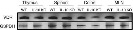
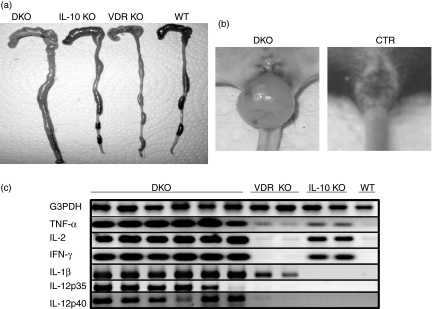
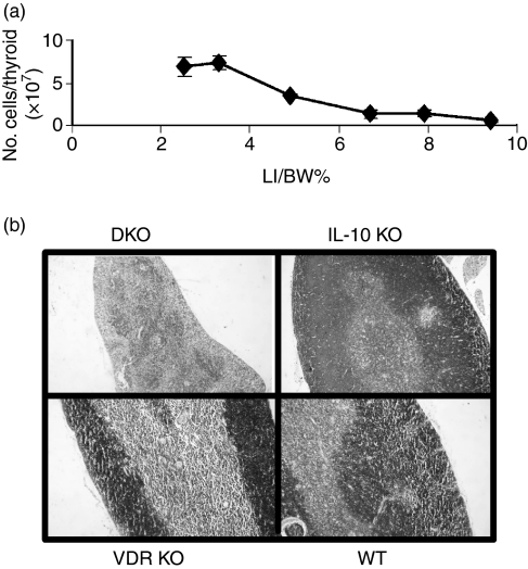
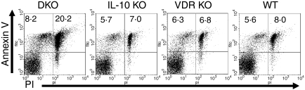
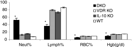
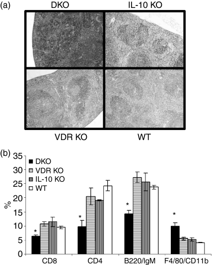
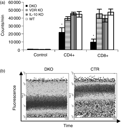
The vitamin D receptor (VDR) is a nuclear receptor expressed in a number of different cells of the immune system. This study was performed to determine the effect of VDR deficiency on immune function and inflammation of the gastrointestinal tract in a model of inflammatory bowel disease, namely interleukin-10 (IL-10) knockout mice. IL-10 knockout mice were generated which either could or could not respond to vitamin D (double IL-10/VDR knockout; DKO). The distribution and function of lymphocytes in both the primary and secondary lymphoid organs were compared and determined as a function of the severity of intestinal inflammation. DKO mice had normal thymic development and peripheral T-cell numbers at 3 weeks of age, but a week after intestinal disease was detected the thymus was dysplastic with a reduction in cellularity. The atrophy was coupled with increased apoptosis. The spleen weight of DKO mice increased as a result of the accumulation of red blood cells; however, there was a 50% reduction in the numbers of T and B cells. Conversely, the mesenteric lymph nodes were enlarged and contained increased numbers of lymphocytes. The T cells from DKO mice were of a memory phenotype and were hyporesponsive to T-cell receptor stimulation. Colitis in the DKO mice was associated with local and high expression of IL-2, interferon-gamma, IL-1beta, tumour necrosis factor-alpha and IL-12. The primary and secondary lymphoid organs in DKO mice are profoundly altered as a consequence of the fulminating inflammation in the gastrointestinal tract. VDR expression is required for the T cells and other immune cells to control inflammation in the IL-10 KO mice.


Figures

Figure 1
Figure 1
Constitutive expression of the VDR in thymus, spleen, MLN and colon of 5-week-old IL-10 KO and WT mice. RNA was isolated, reverse transcribed and then amplified using PCR with primers specific for the VDR and G3PDH. The G3PDH bands indicate that equivalent amounts of mRNA were analysed in each sample. One representative experiment of five is shown.
Figure 2
Figure 2
Fulminant colitis and cytokine expression in the colons of DKO mice. (a) The caecum and colon of DKO, IL-10 KO, VDR KO and WT mice. The DKO colon is enlarged compared to the colons of age-matched single KO or WT mice. (b) Rectal prolapse is common in DKO mice at 5 weeks of age. Age-matched IL-10 KO, VDR KO and WT (CTR-control) mice did not develop colonic prolapse. (c) Cytokine expression in the colon of DKO mice. RT-PCR amplification of G3PDH, TNF-α, IL-2, IFN-γ, IL-1β, IL-12 p35 and IL-12 p40 mRNA extracted from colonic tissue of mice at 5 weeks of age. The PCR products were run on a 1·5% agarose gel and stained with ethidium bromide. Since the G3PDH bands are of approximately equal intensity, changes in the intensities of other bands indicate differences in expression of those genes in those samples.
Figure 3
Figure 3
Thymic involution parallels colitis development in DKO mice. (a) Cell number in the thymus of DKO mice as a function of the ratio of the LI/BW%. (b) H&E staining of the thymus.
Figure 4
Figure 4
Increased thymocyte susceptibility to apoptosis. Thymocytes were stained with annexin V and PI. The percentage of cells that were in early apoptosis (annexin V+ PI) or late apoptosis (annexin V+ PI+) were determined. The results are representative of experiments with six mice per group. DKO values for late apoptosis were significantly different from single KO and WT values. P < 0·05.
Figure 5
Figure 5
Anaemia and alterations in neutrophil and lymphocyte subpopulations in the peripheral blood of DKO mice. Blood was analysed using the Advia system and the percentages of neutrophils (Neut), lymphocytes (Lymph), red blood cells (RBC) and haemoglobin (Hgb) in the blood of each strain were determined. *Data are expressed as mean % ± SE of six mice per group. DKO values were significantly different than those from VDR KO, IL-10 KO and WT mice. P < 0·05.
Figure 6
Figure 6
The spleen of DKO mice is larger and contains more macrophages but fewer T and B cells than the spleens of single KO or WT controls. (a) H&E staining of the whole spleen sections. (b) Single cell suspensions of spleens from DKO, IL-10 KO, VDR KO and WT mice were analysed by flow cytometry for the expression of CD4, CD8, B220/IgM, and F4/80 CD11b. Reduced CD4, CD8 and, B220/IgM proportions and increased F4/80 CD11b proportions in splenocytes from DKO mice. *DKO values were significantly different than the corresponding VDR KO, IL-10 KO and WT values. P < 0·05 n = 6 to n = 12.
Figure 7
Figure 7
Altered proliferation of DKO T cells. (a) Purified CD4+ and CD8+ were isolated from DKO, VDR KO, IL-10 KO and WT mice and stimulated for 72 hr with plate-bound CD3 antibodies. The CD4+ and CD8+ T cells from DKO mice were relatively unresponsive to CD3 stimulation compared to the same cells from VDR KO, IL-10 KO and WT mice. Bars represent means ± SE (n = 5 or n = 6). *P < 0·05. (b) Single cell suspensions of splenocytes from DKO mice were compared to single KO and WT mice for the resting levels of intracellular Ca2+. DKO splenocytes had lower levels of intracellular Ca2+ (dark band at lower fluoresence intensity) compared to VDR KO, IL-10 KO, and WT splenocytes (control, CTR).

References

    1. Gerd Bouma WS. The immunological and genetic basis of inflammatory bowel disease. Nature Rev Immunol. 2003;3:521–33. - PubMed
    1. Podolsky D. Lessons from genetic models of inflammatory bowel disease. Acta Gastroenterol Belg. 1997;60:163–5. - PubMed
    1. Cantorna MT. Vitamin D and autoimmunity: is vitamin D status an environmental factor affecting autoimmune disease prevalence? Proc Soc Exp Biol Med. 2000;223:230–3. - PubMed
    1. Sentongo TA, Semaeo EJ, Stettler N, Piccoli DA, Stallings VA, Zemel BS. Vitamin D status in children, adolescents, and young adults with Crohn disease. Am J Clin Nutr. 2002;76:1077–81. - PubMed
    1. Cantorna MT, Humpal-Winter J, DeLuca HF. In vivo upregulation of interleukin-4 is one mechanism underlying the immunoregulatory effects of 1,25-dihydroxyvitamin D(3) Arch Biochem Biophys. 2000;377:135–8. - PubMed

Publication types

MeSH terms

LinkOut - more resources