. 2006 Mar;117(3):319-28.
doi: 10.1111/j.1365-2567.2005.02303.x.

Luminal bacterial antigen-specific CD4+ T-cell responses in HLA-B27 transgenic rats with chronic colitis are mediated by both major histocompatibility class II and HLA-B27 molecules

Affiliations

Luminal bacterial antigen-specific CD4+ T-cell responses in HLA-B27 transgenic rats with chronic colitis are mediated by both major histocompatibility class II and HLA-B27 molecules

Bi-Feng Qian et al. Immunology. 2006 Mar.

Abstract

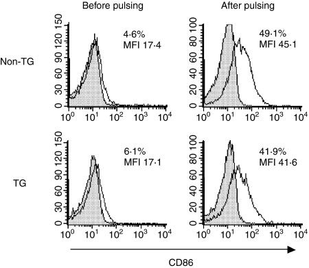
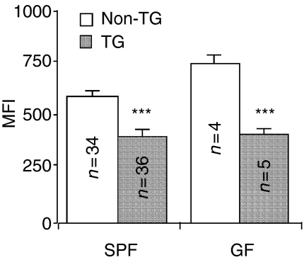
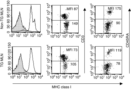
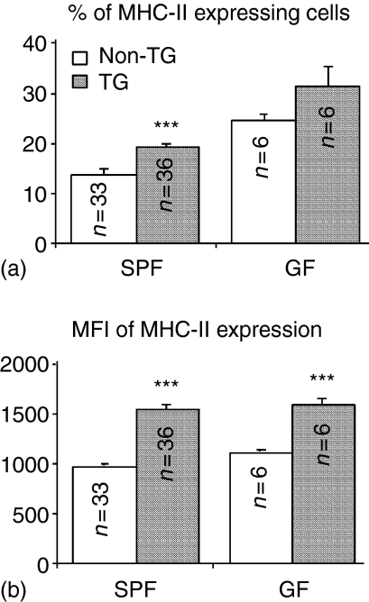
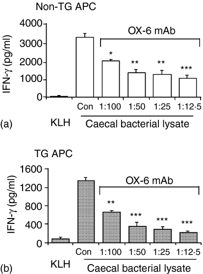
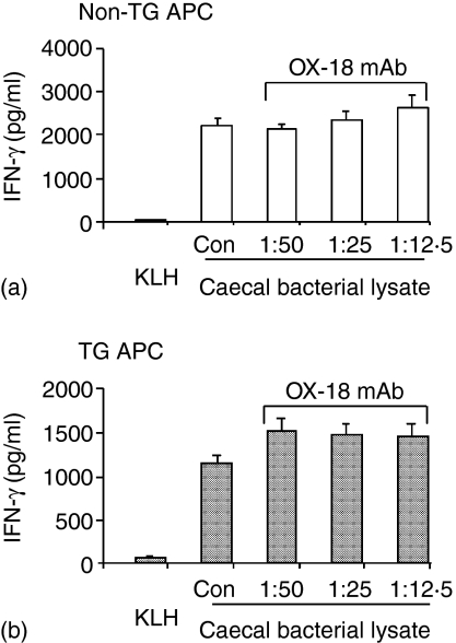
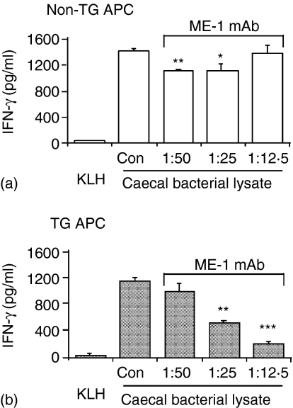
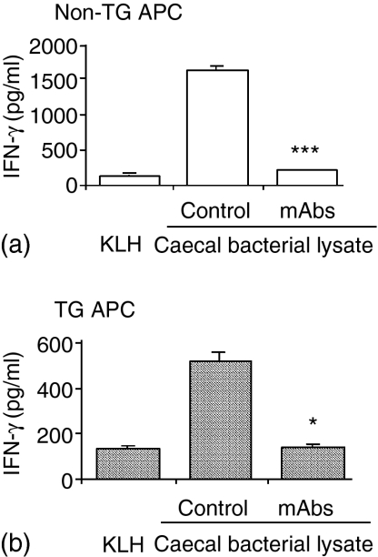
Rats transgenic (TG) for the human major histocompatibility complex (MHC) class I HLA-B27 and beta2-microglobulin genes develop chronic colitis under specific pathogen-free (SPF) but not sterile (germ-free, GF) conditions. We investigated the role of antigen-presenting molecules involved in generating immune responses by CD4+ mesenteric lymph node (MLN) cells from colitic HLA-B27 TG rats to commensal enteric micro-organisms. All TG MLN cells expressed HLA-B27. A higher level of MHC class II was expressed on cells from TG rats, both SPF and GF, compared to non-TG littermates. In contrast, rat MHC class I expression was lower on TG than non-TG cells. Both TG and non-TG antigen presenting cells (APC) pulsed with caecal bacterial antigens induced a marked interferon-gamma (IFN-gamma) response in TG CD4+ T lymphocytes but failed to stimulate non-TG cells. Blocking MHC class II on both TG and non-TG APC dramatically inhibited their ability to induce TG CD4+ T cells to produce IFN-gamma. Blocking HLA-B27 on TG APC similarly inhibited IFN-gamma responses. When the antibodies against MHC class II and HLA-B27 were combined, no APC-dependent IFN-gamma response was detected. These data implicate both native rat MHC class II and TG HLA-B27 in CD4+ MLN T-cell IFN-gamma responses to commensal enteric microflora in this colitis model.


Figures

Figure 1
Figure 1
IFN-γ production by SPF HLA-B27 TG MLN CD4+ T lymphocytes cocultured with caecal bacterial lysate-pulsed SPF or GF APC preparations. Co-culture supernatants were harvested on day 3 and assayed by ELISA. Values represent mean ± SEM of pg/ml of IFN-γ in triplicate culture supernatants. Results shown are representative of three independent experiments. Statistically significant differences between SPF TG and non-TG APC or between SPF and GF TG APC in inducing IFN-γ production by SPF TG CD4+ cells are denoted by asterisks (*P < 0·05, **P < 0·01).
Figure 2
Figure 2
Surface expression of CD86 on T-cell depleted MLN cells from SPF non-TG (upper panels) and TG (lower panels) rats before (left column) and after (right column) overnight pulsing with caecal bacterial lysate. Isotype control staining with FITC labelled mouse IgG1 is shown as filled histograms. Values indicate percentage of positive cells and MFI. Results are representative of three independent experiments.
Figure 3
Figure 3
Analyses of surface expression of rat MHC class I on freshly isolated SPF or GF MLN cells. Unseparated MLN cells were incubated with unlabelled hybridoma cell culture supernatant OX-18. Binding of OX-18 was detected by FITC-labelled goat anti-mouse IgG(γ) antibody. MFI is shown as mean ± SEM. Statistically significant differences between TG and non-TG cells are denoted by asterisks (***P < 0·001). n = number of rats/group.
Figure 4
Figure 4
Cell surface expression of rat MHC class I. Histograms showing MHC class I expression on unseparated MLN cells from SPF non-TG and TG rats, detected by indirect fluorescence using unlabelled OX-18 followed by FITC labelled goat anti-mouse IgG(γ) (black line) compared to control staining with the FITC-labelled secondary antibody alone (filled) are shown in the left column. Dot plots showing MHC class I expression on CD3-positive and CD3-negative MLN cells are in the middle column. Dot plots showing MHC class I expression on CD45RA-positive and CD45RA-negative MLN cells are in the right column. For identification of MHC class I expression on T and B cells, direct fluorescence of FITC-labelled anti-MHC class I monoclonal antibody (OX-18) and PE-anti-CD3 or PE-anti-CD45RA monoclonal antibody was evaluated. Histograms showing all MLN cells are representative of over 30 individual TG and non-TG rats. Dot plots showing two-colour analysis for MHC class I and CD3 or CD45RA are representative of three individual TG and non-TG rats.
Figure 5
Figure 5
Analyses of surface expression of MHC class II (RT1.B detected by OX-6 monoclonal antibody) on freshly isolated MLN cells. The percentage of positive cells and MFI are presented as mean ± SEM. Statistically significant differences between TG and non-TG rats are denoted by asterisks (***P < 0·001). n = number of rats/group.
Figure 6
Figure 6
Blockade of MHC class II significantly inhibited IFN-γ production induced by caecal bacterial lysate-pulsed APC in cocultures of TG CD4+ T lymphocytes plus non-TG APC (a) or TG APC (b). Anti-MHC class II RT1.B antibody (OX-6 monoclonal antibody) at the dilutions indicated, or the isotype control (Con), purified mouse IgG1 at the amount equivalent to the highest concentration of OX-6 mAb, was added to the pulsed SPF APC for 30 min before the start of cocultures with SPF TG CD4+ T lymphocytes. Supernatants were harvested at day 3 and assayed by ELISA. Values represent mean ± SEM of pg/ml of IFN-γ in triplicate culture supernatants. TG CD4+ cells cocultured with an unrelated antigen KLH-pulsed APC served as a control. Results shown are representative of six independent experiments. Statistically significant differences between the amounts of IFN-γ produced in cocultures containing APC treated with anti-MHC class II antibody and in cocultures containing APC treated with the isotype control are denoted by asterisks (*P < 0·05, **P < 0·01, ***P < 0·001).
Figure 7
Figure 7
Blockade of rat MHC class I did not alter IFN-γ production induced by caecal bacterial lysate-pulsed APC in cocultures of TG CD4+ T lymphocytes plus non-TG APC (a) or TG APC (b). Anti-MHC class I antibody (OX-18 monoclonal antibody) at the dilutions indicated, or the isotype control (Con), purified mouse IgG1, at the amount equivalent to the highest concentration of OX-18 mAb, was added to the pulsed SPF APC for 30 min before the start of cocultures with SPF TG CD4+ T lymphocytes. Supernatants were harvested at day 3 and assayed by ELISA. Values represent mean ± SEM of pg/ml of IFN-γ in triplicate culture supernatants. TG CD4+ cells cocultured with an unrelated antigen KLH-pulsed APC served as a control. Results shown are representative of three independent experiments.
Figure 8
Figure 8
Blockade of HLA-B27 markedly inhibited IFN-γ production induced by caecal bacterial lysate-pulsed APC in cocultures with TG CD4+ T lymphocytes. Anti-HLA-B27 antibody (ME-1 monoclonal antibody) at the dilutions indicated, or the isotype control (Con), purified mouse IgG1 at the amount equivalent to the highest concentration of ME-1 monoclonal antibody, was added to the pulsed SPF APC for 30 min before the start of cocultures with SPF TG CD4+ T lymphocytes. Supernatants were harvested at day 3 and assayed by ELISA. Values represent mean ± SEM of pg/ml of IFN-γ in triplicate culture supernatants of TG CD4+ T lymphocytes cocultured with non-TG APC (a) or TG APC (b). TG CD4+ cells cocultured with an unrelated antigen KLH-pulsed APC served as a control. Results shown are representative of six independent experiments. Statistically significant differences between the amounts of IFN-c produced in cocultures containing APC treated with anti-HLA-B27 antibody and in cocultures containing APC treated with isotype control are denoted by asterisks (*P < 0·05, **P < 0·01, ***P < 0·001).
Figure 9
Figure 9
Combined administration of antibodies to MHC class II and HLA-B27 completely inhibited IFN-γ production induced by caecal bacterial lysate-pulsed APC in cocultures with TG CD4+ T lymphocytes. Antibodies were added to the pulsed SPF APC for 30 min before the start of cocultures with SPF TG CD4+ T lymphocytes. The isotype control, purified mouse IgG1, was added at the amount equivalent to the highest concentration of the antibodies used. Supernatants were harvested at day 3 and assayed by ELISA. Values represent mean ± SEM of pg/ml of IFN-γ in triplicate culture supernatants of TG CD4+ T lymphocytes cocultured with (a) non-TG APC in the presence of anti-MHC class II RT1. B (OX-6 monoclonal antibody, dilution 1 : 50) plus anti-MHC class II RT1. D (OX-17 monoclonal antibody, dilution 1 : 250) or (b) TG APC in the presence of anti-MHC class II RT1.B, anti-MHC class II RT1.D, and anti-HLA-B27 (ME-1 monoclonal antibody, dilution 1 : 25). TG CD4+ cells cocultured with an unrelated antigen KLH-pulsed APC served as a control. Results shown are representative of four independent experiments. Statistically significant differences between the APC treated with the specific antibodies and the isotype control in inducing IFN-γ production of TG CD4+ cells are denoted by asterisks (*P < 0·05, ***P < 0·001).

Similar articles

Cited by

References

    1. Sartor RB. Microbial influences in inflammatory bowel disease: role in pathogenesis and clinical implications. In: Sartor RB, Sandborn WJ, editors. Kirsner's Inflammatory Bowel Diseases. Philadelphia: Elsevier; 2004. pp. 138–62.
    1. Sartor RB. Animal models of intestinal inflammation. In: Sartor RB, Sandborn WJ, editors. Kirsner's Inflammatory Bowel Diseases. Philadelphia: Elsevier; 2004. pp. 120–37.
    1. Davidson NJ, Hudak SA, Lesley RE, Menon S, Leach MW, Rennick DM. IL-12, but not IFN-gamma, plays a major role in sustaining the chronic phase of colitis in IL-10-deficient mice. J Immunol. 1998;161:3143–9. - PubMed
    1. Neurath MF, Fuss I, Kelsall BL, Stuber E, Strober W. Antibodies to interleukin 12 abrogate established experimental colitis in mice. J Exp Med. 1995;182:1281–90. - PMC - PubMed
    1. Powrie F, Leach MW, Mauze S, Menon S, Caddle LB, Coffman RL. Inhibition of Th1 responses prevents inflammatory bowel disease in scid mice reconstituted with CD45RBhi CD4+ T cells. Immunity. 1994;1:553–62. - PubMed

Publication types

MeSH terms