. 2006 Mar;117(3):329-39.
doi: 10.1111/j.1365-2567.2005.02304.x.

Penetratin tandemly linked to a CTL peptide induces anti-tumour T-cell responses via a cross-presentation pathway

Affiliations

Penetratin tandemly linked to a CTL peptide induces anti-tumour T-cell responses via a cross-presentation pathway

Dodie S Pouniotis et al. Immunology. 2006 Mar.

Abstract

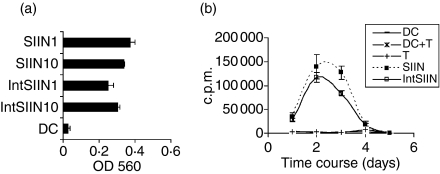
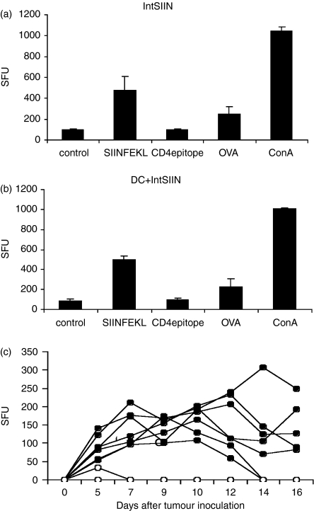
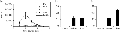
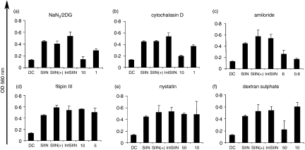
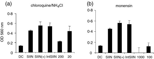
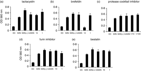
Recently there has been increasing evidence to suggest that membrane translocating peptides enter cells by a receptor-dependent pathway. There have been some studies on the mechanism of major histocompatibility complex (MHC) class I presentation of membrane translocating peptides incorporating cytotoxic T lymphocyte epitopes. However, these have been on different cell lines and only a limited number of inhibitors of the antigen presentation pathway were used. Herein, we demonstrate a comprehensive study utilizing a full spectrum of inhibitors to various pathways of MHC class I to elucidate the mechanism of the membrane translocating peptide, penetratin from Antennapedia (Int). It is clear that Int, RQIKIWFQNRRMKWKK when tandemly linked to a cytotoxic T lymphocyte peptide of ovalbumin, SIINFEKL (IntSIIN) is endocytosed via phagocytosis or macropinocytosis by dendritic cells in an ATP-dependent manner and is processed by a proteasome- and tapasin-independent pathway for presentation and loading to MHC class I molecules. In addition, the majority of antigen is taken up by negatively charged receptors. IntSIIN activates T cells in vitro and in vivo and protects mice against challenge with an ovalbumin-expressing tumour.


Figures

Figure 1
Figure 1
IntSIIN stimulates T cells in vitro. (a) C57BL/6 DC were stimulated with IntSIIN and SIIN at 10 and 1 μg/ml for 1 hr and added to B3Z T cells for 24 hr. LacZ activity in B3Z T cells was assayed by total culture lysates with LacZ substrate CPRG. The absorbance (560 nm) of culture wells was read after 4 hr incubation at 37°. (b) C57BL/6 DC were pulsed with SIIN and IntSIIN at 1 μg/ml for 1 hr, washed, and added to purified OT-I T cells and cultured for 6 days. Cells were harvested at each time-point and [3H]thymidine uptake was measured to assess T-cell proliferation. DC alone, OT-I T cells and DC added to T cells were used in experiments as negative controls and are shown in both (a) and (b). Data are presented as counts per minute (c.p.m.) of [3H]thymidine uptake from quadruple wells (± SD).
Figure 2
Figure 2
IFN-γ responses and tumour protection in C57BL/6 mice immunized with IntSIIN. C57BL/6 mice were immunized twice at day 0 and 14 with (a)IntSIIN and (b) DC pulsed with IntSIIN. Immunizations were given intradermally and 16 days post-immunization, spleens were removed for ELISpot analysis. IFN-γ responses are given as spot-forming units (SFU) per 0·5 million cells. Representative results from one of three experiments are shown. One or two immunizations did not show any difference in responses; presented here after one injection. (c) Groups of C57BL/6 mice received one injection intradermally with (closed circles) PBS alone and (open circles) IntSIIN at 25 μg per mouse. After 14 days mice received a subcutaneous challenge of 107 E.G7.OVA tumour cells. Individual mouse curves are shown (n = 7).
Figure 3
Figure 3
TAP-independent stimulation of CD8 T-cell responses by IntSIIN. (a) In vitro grown TAP−/− DC were pulsed with SIIN and IntSIIN at 1 μg/ml for 1 hr then added to purified OT-I T cells and cultured for 6 days. Cells were harvested at each time-point and uptake of [3H]thymidine was measured to assess T-cell proliferation. DC pulsed with nothing, OT-I T cells and DC added to T cells were used in experiments as negative controls. Data are presented as counts per minute (c. p.m.) from quadruple wells (± SD). (b) EL-4 and (c) RMA-S were stimulated with IntSIIN and SIIN at 1 μg/ml for 1 hr and added to B3Z T cells for 24 hr. LacZ activity in B3Z T cells was assayed by total culture lysates with LacZ substrate CPRG. EL-4 and RMA-S added to B3Z T cells without peptides were used in experiments as negative controls. The absorbance (560 nm) of culture wells was read after 4 hr incubation at 37°.
Figure 4
Figure 4
Uptake of IntSIIN. DC were incubated for 45 min with inhibitors (a) sodium azide/2-deoxyglucose (10, 1 mm), (b) cytochalasin D (10, 1 μg/ml), (c) amiloride (6, 0·6 μm), (d) filipin III (10, 1 μg/ml), (e) nystatin (50, 10 μg/ml) and (f) dextran sulphate (50, 10 μg/ml), followed by incubation with IntSIIN at 1 μg/ml and added to B3Z T cells for 24 hr. DC were pulsed with SIINFEKL in the presence of the biochemical inhibitor at the maximum dose and were added to B3Z T cells in parallel experiments to ensure no inhibitory effects on the presentation of SIINFEKL CTL epitope. LacZ activity in B3Z T cells was assayed by total culture lysates with LacZ substrate CPRG. The absorbance (560 nm) of culture wells was read after 4 hr incubation at 37°.
Figure 5
Figure 5
IntSIIN is endocytosed and processed in endosomes. DC were incubated for 45 min with inhibitors (a) chloroquine/NH4CL (200, 20 μm), and (b) monensin (1000, 100 μm) followed by incubation with IntSIIN at 1 μg/ml and added to B3Z T cells for 24 hr. DC were pulsed with SIINFEKL in the presence of the biochemical inhibitor at the maximum dose and added to B3Z T cells in parallel experiments to ensure no inhibitory effects on the presentation of SIINFEKL CTL epitope. LacZ activity in B3Z T cells was assayed by total culture lysates with LacZ substrate CPRG. The absorbance (560 nm) of culture wells was read after 4 hr incubation at 37°.
Figure 6
Figure 6
Proteolysis and peptide loading of IntSIIN. DC were incubated for 45 min with inhibitors, (a) lactacystin (10, 1 μm), (b) brefeldin (10, 1 μg/ml), (c) protease inhibitor (1/100, 1/10 dilution), (d) furin inhibitor (10, 1 μm) and (e) bestatin (10, 1 μm) followed by incubation with IntSIIN at 1 μg/ml and added to B3Z T-cell hybridoma for 24 hr. DC were pulsed with SIINFEKL in the presence of the biochemical inhibitor at the maximum dose and added to B3Z T cells in parallel experiments to ensure no inhibitory effects on the presentation of SIINFEKL CTL epitope. LacZ activity in B3Z T cells was assayed by total culture lysates with LacZ substrate CPRG. The absorbance (560 nm) of culture wells was read after 4 hr incubation at 37°.

Similar articles

Cited by

References

    1. Pietersz GA, Apostolopoulos V, McKenzie IF. Generation of cellular immune responses to antigenic tumor peptides. Cell Mol Life Sci. 2000;57:290–310. - PMC - PubMed
    1. Trombetta ES, Mellman I. Cell biology of antigen processing in vitro and in vivo. Annu Rev Immunol. 2005;23:975–1028. - PubMed
    1. Apostolopoulos V, Barnes N, Pietersz GA, McKenzie IF. Ex vivo targeting of the macrophage mannose receptor generates anti-tumor CTL responses. Vaccine. 2000;18:3174–84. - PubMed
    1. Apostolopoulos V, Lofthouse SA, Popovski V, Chelvanayagam G, Sandrin MS, McKenzie IF. Peptide mimics of a tumor antigen induce functional cytotoxic T cells. Nat Biotechnol. 1998;16:276–80. - PubMed
    1. Apostolopoulos V, McKenzie IF. Cellular mucins: targets for immunotherapy. Crit Rev Immunol. 1994;14:293–309. - PubMed

Publication types

MeSH terms