

Idiopathic sclerosing encapsulating peritonitis (or abdominal cocoon)
- PMID: 16476161
- PMCID: PMC1421435
- DOI: 10.1186/1471-2482-6-3
Idiopathic sclerosing encapsulating peritonitis (or abdominal cocoon)
Abstract
Background: Idiopathic sclerosing encapsulating peritonitis (or abdominal cocoon) is a rare cause of small bowel obstruction, especially in adult population. Diagnosis is usually incidental at laparotomy. We discuss one such rare case, outlining the fact that an intra-operative surprise diagnosis could have been facilitated by previous investigations.
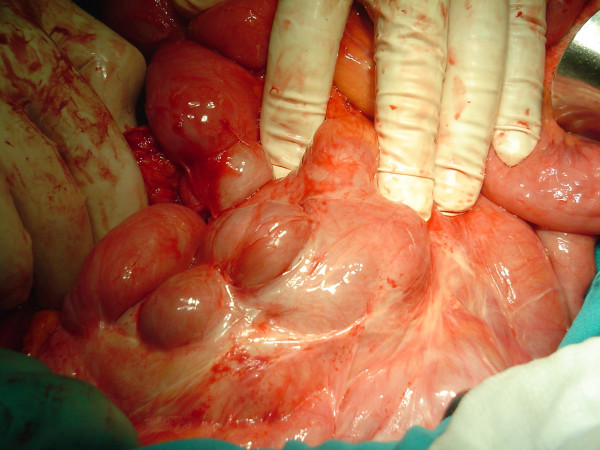
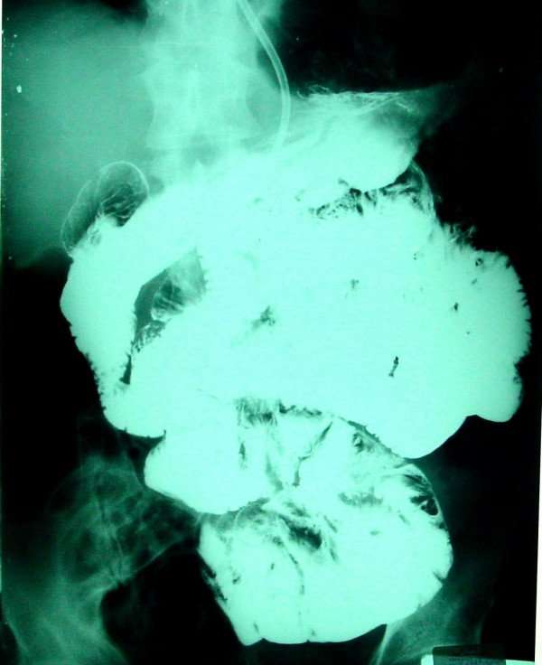
Case presentation: A 56 year-old man presented in A&E department with small bowel ileus. He had a history of 6 similar episodes of small bowel obstruction in the past 4 years, which resolved with conservative treatment. Pre-operative work-up did not reveal any specific etiology. At laparotomy, a fibrous capsule was revealed, in which small bowel loops were encased, with the presence of interloop adhesions. A diagnosis of abdominal cocoon was established and extensive adhesiolysis was performed. The patient had an uneventful recovery and follow-up.
Conclusion: Idiopathic sclerosing encapsulating peritonitis, although rare, may be the cause of a common surgical emergency such as small bowel ileus, especially in cases with attacks of non-strangulating obstruction in the same individual. A high index of clinical suspicion may be generated by the recurrent character of small bowel ileus combined with relevant imaging findings and lack of other plausible etiologies. Clinicians must rigorously pursue a preoperative diagnosis, as it may prevent a "surprise" upon laparotomy and result in proper management.
Figures
References
-
- Foo KT, Ng KC, Rauff A, Foong WC, Sinniah R. Unusual small intestinal obstruction in adolescent girls: the abdominal cocoon. Br J Surg. 1978;65:427–430. - PubMed
-
- Kawaguchi Y, Kawanishi H, Mujais S, Topley N, Oreopoulos DG. Encapsulating peritoneal sclerosis: definition, etiology, diagnosis, and treatment. International Society for Peritoneal Dialysis Ad Hoc Committee on Ultrafiltration Management in Peritoneal Dialysis. Perit Dial Int. 2000;20 Suppl 4:S43–55. - PubMed
-
- Hur J, Kim KW, Park MS, Yu JS. Abdominal cocoon: preoperative diagnostic clues from radiologic imaging with pathologic correlation. AJR Am J Roentgenol. 2004;182:639–641. - PubMed
Publication types
MeSH terms
LinkOut - more resources
Full Text Sources
Miscellaneous
