. 2006 Feb 13:4:2.
doi: 10.1186/1477-3155-4-2.

Canine parvovirus-like particles, a novel nanomaterial for tumor targeting

Affiliations

Canine parvovirus-like particles, a novel nanomaterial for tumor targeting

Pratik Singh et al. J Nanobiotechnology. .

Abstract

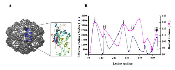
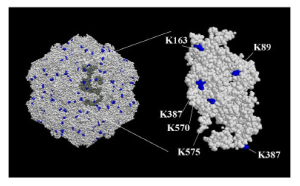
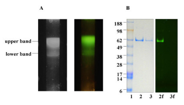
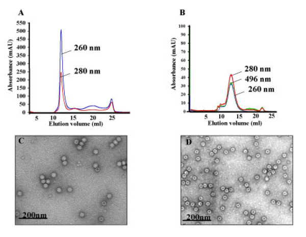
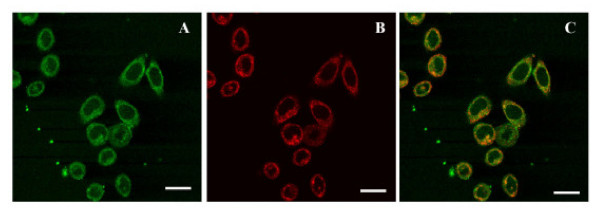
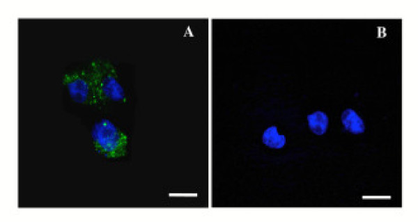
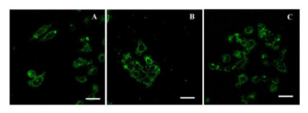
Specific targeting of tumor cells is an important goal for the design of nanotherapeutics for the treatment of cancer. Recently, viruses have been explored as nano-containers for specific targeting applications, however these systems typically require modification of the virus surface using chemical or genetic means to achieve tumor-specific delivery. Interestingly, there exists a subset of viruses with natural affinity for receptors on tumor cells that could be exploited for nanotechnology applications. For example, the canine parvovirus (CPV) utilizes transferrin receptors (TfRs) for binding and cell entry into canine as well as human cells. TfRs are over-expressed by a variety of tumor cells and are widely being investigated for tumor-targeted drug delivery. We explored whether the natural tropism of CPV to TfRs could be harnessed for targeting tumor cells. Towards this goal, CPV virus-like particles (VLPs) produced by expression of the CPV-VP2 capsid protein in a baculovirus expression system were examined for attachment of small molecules and delivery to tumor cells. Structural modeling suggested that six lysines per VP2 subunit are presumably addressable for bioconjugation on the CPV capsid exterior. Between 45 and 100 of the possible 360 lysines/particle could be routinely derivatized with dye molecules depending on the conjugation conditions. Dye conjugation also demonstrated that the CPV-VLPs could withstand conditions for chemical modification on lysines. Attachment of fluorescent dyes neither impaired binding to the TfRs nor affected internalization of the 26 nm-sized VLPs into several human tumor cell lines. CPV-VLPs therefore exhibit highly favorable characteristics for development as a novel nanomaterial for tumor targeting.


Figures

Figure 1
Figure 1
CPV Capsid and subunit organization. The 2CAS model of CPV was downloaded from the VIPER database. The expanded inset shows a single VP2 subunit ribbon diagram with N-terminus in blue and C-terminus in red. B. Accessible surface lysines profile of CPV capsid. Data shown was downloaded from VIPER database. Lysine residues in the VP2 subunit are shown on X-axis (total of 20 per subunit) and the effective radius multiplied by the solvent accessible surface area (SASA) is shown in blue on Y-axis on the left side. The radial distance of each residue is also shown on Y-axis in magenta. Coinciding high values on Y-axis suggest residues that are (i) highly accessible, (ii) moderately accessible and (iii) accessible to a lesser extent.
Figure 2
Figure 2
Space filling model of surface accessible lysines on CPV capsid. The CPV capsid model was generated with VMD software. The figure shows identified accessible lysines on CPV based upon the whole capsid (left side) and on an individual VP2 subunit (right side).
Figure 3
Figure 3
CPV purification and characterization. A. Sucrose gradient purification. VLPs preparation from infected cell culture lysates purified by sucrose gradient centrifugation (10–40%). Bands of CPV-VLPs that were derivatized with OG-488 are visible in the gradient just above the middle of the tube (left panel) and appear fluorescent green under a UV-light source (right panel). B. SDS-PAGE analyses. The purified VLPs were subjected to electrophoresis in 4–12% Bis-tris gel and stained with SimplyBlue (Invitrogen) to reveal the proteins (left panel). The Seeblue plus protein molecular weight standards in kDa (Invitrogen) are indicated on the side of the gel picture (lane 1). Lanes 2 and 3 contain protein from CPV-VLPs derivatized with OG-488 and CPV-VLPs respectively. Prior to staining, the gel (right panel) visualized with a UV-light source showed a fluorescent 62 kDa band in the lane of OG-488 derivatized CPV-VLPs (lane 2f) and lacked any fluorescent bands in the native CPV-VLPs (lane 3f).
Figure 4
Figure 4
Capsid stability and morphology of CPV-VLPs. A and B. Size exclusion chromatography (SEC) of CPV-VLPs. Sucrose gradient purified samples were passed through a Superose6 size exclusion column. Absorbance values recorded at 260 nm (for nucleic acids), 280 nm (for protein) and 496 nm (for OG-488 dye) are shown on the y-axis. The elution profile from the column in ml is shown on x-axis. Panel A shows SEC of freshly purified CPV-VLPs and panel B shows SEC of CPV-VLPs labeled with OG-488 dye following 1 week of storage at 4°C. C and D. Electron micrographs of CPV-VLPs. CPV-VLPs were deposited onto carbon-coated copper grids and stained with uranyl acetate. The micrographs of (C) full capsids in a freshly purified CPV-VLPs preparation and (D) empty capsids in CPV-VLPs sample after 1 week of storage are shown. Both micrographs were taken at a nominal magnification of 60,000×.
Figure 5
Figure 5
Binding and internalization into of CPV-VLPs into HeLa cells. HeLa cells incubated with Texas red-labeled transferrin (red) and CPV-VLPs were washed and fixed. Labeled antibodies (green) were used to detect the presence of CPV-VLPs in the cells by fluorescence confocal microscopy. (A) CPV-VLPs are seen as green areas in the cytoplasm, (B) shows localization of Texas Red-transferrin (red) and (C) depicts merged picture showing co-localization of CPV-VLPs and transferrin in yellow. Scale bar, 25 μm.
Figure 6
Figure 6
Binding and internalization of CPV-VLPs labeled with OG-488 into transferrin receptor expressing cells. Cells differing in level of transferrin receptor expression, TRVb1 (express TfRs) and TRVb (low or lacking TfR expression) were exposed to CPV-VLPs. Internalized dye-labeled CPV-VLPs were detected by fluorescence confocal microscopy. TOTO-3 (blue) was used for staining the nuclei. (A) TRVb1 cells with internalized dye-derivatized CPV-VLPs, are seen as green areas in the cytoplasm, and (B) TRVb cells show lack of VLP internalization. Scale bar, 25 μm.
Figure 7
Figure 7
Binding and internalization of CPV-VLPs labeled with OG-488 into tumor cell lines. Tumor cell lines (A) HeLa, (B) HT-29 and (C) MDA-MB231 were exposed to OG488-labeled CVP-VLPs. The cells were washed, fixed and examined by confocal fluorescence microscopy for internalization of the particles. Scale bar, 25 μm.

References

    1. Fennelly D. Dose intensity in advanced ovarian cancer: have we answered the question? Clin Cancer Res. 1995;1:575–582. - PubMed
    1. Myers CE, Chabner BA. Anthracyclins. In: Chabner, B. A., Collins, J. M., editor. Cancer chemotherapy-principles and practice. Philadelphia, Lippincott; 1990. pp. 256–381.
    1. Dubowchik GM, Walker MA. Receptor-mediated and enzyme-dependent targeting of cytotoxic anticancer drugs. Pharmacol Ther. 1999;83:67–123. doi: 10.1016/S0163-7258(99)00018-2. - DOI - PubMed
    1. Feng SS, Chien S. Chemotherapeutic engineering: application and further development of chemical engineering principles for chemotherapy of cancer and other diseases. Chemical Engineering Science. 2003;58:4087–4114. doi: 10.1016/S0009-2509(03)00234-3. - DOI
    1. Brannon-Peppas L, Blanchette JO. Nanoparticle and targeted systems for cancer therapy. Advanced drug delivery reviews. 2004;56:1649–1659. doi: 10.1016/j.addr.2004.02.014. - DOI - PubMed

LinkOut - more resources