. 2006 Feb 20;203(2):275-80.
doi: 10.1084/jem.20051951. Epub 2006 Feb 13.

Human antibodies induce arthritis in mice deficient in the low-affinity inhibitory IgG receptor Fc gamma RIIB

Affiliations

Human antibodies induce arthritis in mice deficient in the low-affinity inhibitory IgG receptor Fc gamma RIIB

Stefka B Petkova et al. J Exp Med. .

Abstract

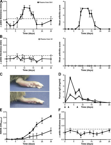
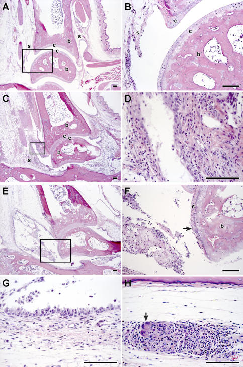
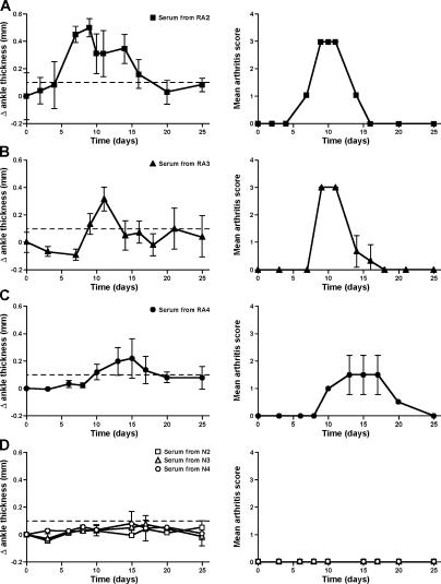
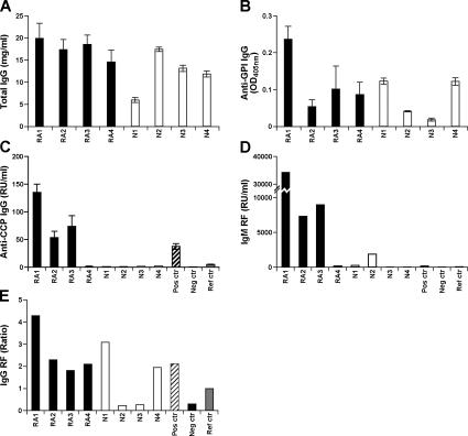
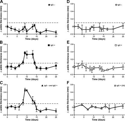
Rheumatoid arthritis (RA) is a complex autoimmune disease with a poorly understood pathogenesis. The disease is associated with polyclonal B cell activation and the production of autoantibodies (autoAbs), but there is a longstanding controversy as to whether such Abs contribute to, or are secondary to, the pathogenesis of RA. To address the potential pathogenicity of human RA-associated Abs, we developed a passive transfer model involving mice deficient in the low-affinity inhibitory Fc receptor, FcgammaRIIB. We report that plasma or serum from patients with active RA can induce inflammation and histological lesions in FcgammaRIIB-/- mice consistent with arthritis, and that this pathogenic activity is caused by the immunoglobulin G-rich fraction. Our results suggest that humoral autoimmunity can contribute directly to autoimmune arthritis, and that FcgammaRIIB-/- mice are a promising model to evaluate the arthritogenic potential of human autoAbs.


Figures

Figure 1.
Figure 1.
Induction of inflammatory arthritis in FcγRIIB−/− with plasma from an RA patient. 8–12-wk-old B6.FcγRIIB−/− mice were injected with plasma from a patient with RA (RA1) or a healthy blood donor (N1) on days 0, 2, and 7. Arthritis was evaluated by measuring the increase in ankle thickness and arthritis score (see Materials and methods). Each solid line represents the mean ± SEM (mm) results from both ankles. Values above the dashed line are considered statistically significant based on measuring the ankle thickness of an untreated control group of B6.FcγRIIB−/− mice. (A) B6.FcγRIIB−/− (n = 6) mice were injected with plasma from RA1, and the animals were examined for a period of 25 d after the initial injection (♦). (B) B6.FcγRIIB−/− mice (n = 4) injected with plasma from a healthy blood donor N1 (⋄). (C) Representative images of hind paws at day 14 after the initial injection with plasma from RA1 (top) and N1 (bottom). (D) Clearance of human IgG Abs in the mouse circulation during the time course of the experiment. Human blood samples were injected on days 0, 2, and 4 as indicated by arrowheads. (E) Accumulation of MAHAs was more pronounced in recipients of plasma from RA1 as compared with control individual N1. (F) B6.FcγRIIB+/+ mice (n = 3) injected with plasma from RA1 did not develop arthritis when injected with the same volume (2.5 ml/mouse) of arthritogenic RA1 plasma.
Figure 2.
Figure 2.
Histopathology of ankle joints. (A) Hematoxylin and eosinstained tissue sections from the ankle joint obtained from a representative B6.FcγRIIB−/− mouse injected with plasma collected from healthy individual N1 and killed on day 11. For orientation: s, synovium; b, bone; c, cartilage. (B) Higher magnification of selected area from A showing a normal joint space, smooth arthricular cartilage, and normal synovial membrane. (C) Section of ankle joint from a mouse injected with plasma from RA patient RA1 and killed on day 11 after the initial injection. Note the marked expansion of the synovial stroma by inflammatory cells. (D) Higher magnification of the synovium shows the infiltration with inflammatory cells is predominantly neutrophilic. (E–H) Representative images of the ankle joint from mice injected with plasma from RA1 and killed on days 14 and 15. (E) Inflammation and synovial hypertrophy are seen on day 14, which coincides with the peak of ankle swelling. (F) Higher magnification of a field from E showing fibrillation of the cartilage surface (arrow) and mixed inflammatory cells, predominantly neutrophils, and macrophages within the joint space. (G) Mild focal synovial hyperplasia. (H) Periarticular soft tissue with perivascular inflammation predominantly containing neutrophils, macrophages, and a few Langerhans-type multinucleated giant cells (arrow). Bar, 100 μm.
Figure 3.
Figure 3.
Passive transfer of arthritis with sera obtained from three additional RA patients. FcγRIIB−/− mice were injected with sera from three additional arthritic patients (RA2, RA3, and RA4; A, B, and C) and three normal blood donors (N2, N3, and N4; D). Ankle thickening (mm) ± SEM and arthritis score are shown. Results for each group of mice were calculated as an average based on individual measurements of both hind paws of each individual mouse. The severity and onset of arthritis after injection with human RA samples varied among recipient groups; however, a 100% incidence was observed in each group (A, n = 2; B, n = 4; C, n = 2). None of the animals injected with sera from normal blood donors (A, n = 1; B, n = 3; C, n = 3) developed ankle swelling.
Figure 4.
Figure 4.
Levels of human IgG Abs. (A) Total IgG (mg/ml). (B) Anti-GPI Abs (OD, 405 nm). (C) Anti-CCP IgG Abs (relative units, RU/ml). Values >5 RU/ml were considered positive. Mean ± SD is indicated. (D) IgM RF (values ≥20 RU/ml are considered positive). (E) IgG RF of each blood sample obtained from RA patients and healthy controls. Data indicate the calculated ratios, and values >1 were considered positive.
Figure 5.
Figure 5.
Induction of arthritis with purified human IgG Abs from patient RA1. Groups of FcγRIIB−/− mice (n = 3 each group) were injected with protein G column-separated IgG and IgG+ fractions of human blood samples. (A, B, and C) IgG (<0.0001 mg IgG/mouse), IgG+ (total 47.4 mg IgG/mouse), and recombined IgG plus IgG+ (47.4 mg IgG/mouse) fractions from RA1 were tested for their ability to induce arthritis in FcγRIIB−/− mice. Arthritis was monitored as ankle thickening (presented here) and the overall arthritis score (not depicted). (D, E, and F) IgG (<0.0001 mg IgG/mouse), IgG+ (14.7 mg IgG/mouse), and 3× IgG+ (44.1 mg IgG/mouse) fractions obtained from N1 were injected in animals, and arthritis was monitored for 25 d.

Similar articles

Cited by

References

    1. Feldmann, M., F.M. Brennan, and R.N. Maini. 1996. Rheumatoid arthritis. Cell. 85:307–310. - PubMed
    1. Dorner, T., K. Egerer, E. Feist, and G.R. Burmester. 2004. Rheumatoid factor revisited. Curr. Opin. Rheumatol. 16:246–253. - PubMed
    1. Schaller, M., W. Stohl, S.M. Tan, V.M. Benoit, D.M. Hilbert, and H.J. Ditzel. 2005. Raised levels of anti-glucose-6-phosaphate isomerase IgG in serum and synovial fluid from patients with inflammatory arthritis. Ann. Rheum. Dis. 64:743–749. - PMC - PubMed
    1. Matsumoto, I., D.M. Lee, R. Goldbach-Mansky, T. Sumida, C.A. Hitchon, P.H. Schur, R.J. Anderson, J.S. Coblyn, M.E. Weinblatt, M. Brenner, et al. 2003. Low prevalence of antibodies to glucose-6-phosphate isomerase in patients with rheumatoid arthritis and a spectrum of other chronic autoimmune disorders. Arthritis Rheum. 48:944–954. - PubMed
    1. Schellekens, G.A., B.A. de Jong, F.H. van den Hoogen, L.B. van de Putte, and W.J. van Venrooij. 1998. Citrulline is an essential constituent of antigenic determinants recognized by rheumatoid arthritis-specific autoantibodies. J. Clin. Invest. 101:273–281. - PMC - PubMed

Publication types