. 2006 Feb 20;203(2):319-23.
doi: 10.1084/jem.20052227. Epub 2006 Feb 13.

Strand-biased defect in C/G transversions in hypermutating immunoglobulin genes in Rev1-deficient mice

Affiliations

Strand-biased defect in C/G transversions in hypermutating immunoglobulin genes in Rev1-deficient mice

Jacob G Jansen et al. J Exp Med. .

Abstract

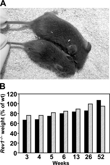
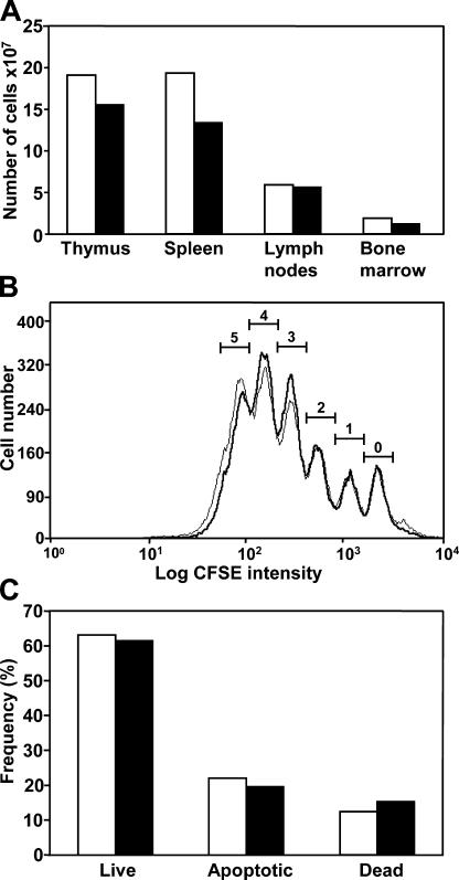
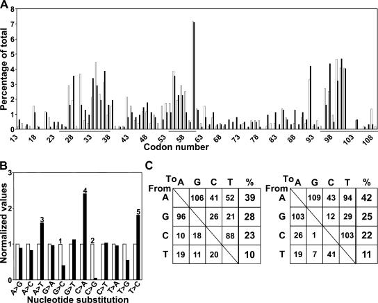
Somatic hypermutation of Ig genes enables B cells of the germinal center to generate high-affinity immunoglobulin variants. Key intermediates in somatic hypermutation are deoxyuridine lesions, introduced by activation-induced cytidine deaminase. These lesions can be processed further to abasic sites by uracil DNA glycosylase. Mutagenic replication of deoxyuridine, or of its abasic derivative, by translesion synthesis polymerases is hypothesized to underlie somatic hypermutation. Rev1 is a translesion synthesis polymerase that in vitro incorporates uniquely deoxycytidine opposite deoxyuridine and abasic residues. To investigate a role of Rev1 in mammalian somatic hypermutation we have generated mice deficient for Rev1. Although Rev1-/- mice display transient growth retardation, proliferation of Rev1-/- LPS-stimulated B cells is indistinguishable from wild-type cells. In mutated Ig genes from Rev1-/- mice, C to G transversions were virtually absent in the nontranscribed (coding) strand and reduced in the transcribed strand. This defect is associated with an increase of A to T, C to A, and T to C substitutions. These results indicate that Rev1 incorporates deoxycytidine residues, most likely opposite abasic nucleotides, during somatic hypermutation. In addition, loss of Rev1 causes compensatory increase in mutagenesis by other translesion synthesis polymerases.


Figures

Figure 1.
Figure 1.
Targeted disruption of Rev1. (A, top) the genomic region of Rev1 that encodes the catalytic domain of Rev1. Vertical bold sections denote exons. SCDE, exon 10 encoding the catalytic domain of Rev1. Horizontal bold sections denote regions homologous to the targeting vector. (A, middle) Targeting vector pSCDE-hygro to delete exon 10. Hyg, pPGK-hygromycine cassette; TK, pPGK-thymidine kinase cassette. Arrows indicate direction of transcription. (A, bottom) Targeted Rev1 allele. Probes A and B, DNA fragments used to analyze gene-targeting events. Ba, BamHI; Bg, BglII; K, KpnI; N, NcoI. (B) Southern blot of genomic mouse DNA digested with KpnI and hybridized with probe A. Fragment sizes indicate the following alleles: 22 kbp, wild type; 11 kbp, Rev1−/−. 1, Rev1+/+; 2, Rev1+/−; 3, Rev1−/−. (C) Western blot of MEF lysates hybridized with an α-Rev1 antiserum. Arrow indicates position of Rev1. 1, Rev1+/+; 2, Rev1−/−. (D) PCR products of Rev1 cDNA amplified from Rev1−/− kidneys. PCR products of exons 10–15 (left) and exons 8–15 (right). 1, size marker; 2, Rev1+/+; 3, Rev1−/−. In the left panel, in lane 3 no PCR product is detected as a consequence of the deletion of exon 10 in the mutant.
Figure 2.
Figure 2.
Retarded growth of Rev1−/− mice. (A) 3-wk-old wild-type (top) and Rev1−/− (bottom) littermates illustrating the reduced body size in young Rev1−/− mice. (B) Growth characteristics of Rev1−/− mice between 3 and 52 wk after birth, expressed as the percentage of weight of wild-type mice. Males (solid bars); females (hatched bars). Each data point represents the mean of 7–10 mice.
Figure 3.
Figure 3.
Cellularity, proliferation, and survival of Rev1−/− B cells. (A) Cellularity of primary and secondary lymphoid organs of wild-type and Rev1−/− mice. At the age of 7 wk, the cellularity of primary and secondary lymphoid organs of wild-type and Rev1−/− mice were determined. The moderate reduction in cellularity likely is related to the reduced size of Rev1−/− mice in comparison with wild-type mice, rather than to a proliferation defect. See Fig. 2 A and Fig. 4 A. (B) Proliferation of CFSE-loaded B cells from wild-type (thin line) and Rev1 mutant mice (bold line). As analyzed by flow cytometry, LPS blasts proliferate normally in the absence of Rev1. (C) Survival of LPS-activated B cells. Live, apoptotic, and dead cells were distinguished by annexinV and propidium iodine (PI) using flow cytometry. Survival and cell death are not significantly influenced by Rev1 under these conditions. Wild type (white bars); Rev1−/− (black bars).
Figure 4.
Figure 4.
Somatic hypermutation of hypermutated Vλ1 genes from memory B cells of wild-type and Rev1−/− mice. (A) Distribution of point mutations in Rev1-deficient B cells. No significant differences are found between both genotypes. Wild-type mice (white bars); Rev1−/− mice (black bars). Mutations concentrate in CDR 1, 2, and 3 (underlined). Numbering of the codons is according to Kabat and Wu (22). (B) Nucleotide substitution profile (normalized to wild-type controls) of the hypermutated Vλ1 segments of the Igλ1 gene derived from Rev1−/− memory B cells. Highly significant differences are found between both genotypes. Wild-type mice (white bars); Rev1−/− mice (black bars). Although the frequency of C to G and, to a lesser extent, G to C transversions are decreased, A to T and C to A transversions as well as T to C transitions are increased. Significant changes (Chi square test) are marked by 1, 2, 3, 4, and 5. The p-values are 0.006, 2 × 10⁻⁵, 0.005, 0.02, and 0.03, respectively. (C) Nucleotide substitution profile in hypermutated Vλ1 sequences from Rev1-proficient and -deficient B cells. The relative contribution of substitutions at all four basepairs is unaltered indicating no overall defect in SHM. (Left) Wild type, n = 508 mutations. (Right) Rev1−/−, n = 587 mutations.

References

    1. Prakash, S., R.E. Johnson, and L. Prakash. 2005. Eukaryotic translesion synthesis DNA polymerases: specificity of structure and function. Annu. Rev. Biochem. 74:317–353. - PubMed
    1. Masuda, Y., M. Takahashi, S. Fukuda, M. Sumii, and K. Kamiya. 2002. Mechanisms of dCMP transferase reactions catalyzed by mouse Rev1 protein. J. Biol. Chem. 277:3040–3046. - PubMed
    1. Zhang, Y., X. Wu, O. Rechkoblit, N.E. Geacintov, J.S. Taylor, and Z. Wang. 2002. Response of human REV1 to different DNA damage: preferential dCMP insertion opposite the lesion. Nucleic Acids Res. 30:1630–1638. - PMC - PubMed
    1. Gibbs, P.E.M., J. McDonald, R. Woodgate, and C.W. Lawrence. 2005. The relative roles in vivo of Saccharomyces cerevisiae Pol eta, Pol zeta, Rev1 protein and Pol32 in the bypass and mutation induction of an abasic site, T-T (6-4) photoadduct and T-T cis-syn cyclobutane dimer. Genetics. 169:575–582. - PMC - PubMed
    1. Simpson, L.J., and J.E. Sale. 2003. Rev1 is essential for DNA damage tolerance and non-templated immunoglobulin gene mutation in a vertebrate cell line. EMBO J. 22:1654–1664. - PMC - PubMed

Publication types

MeSH terms