. 2006 Feb 13;172(4):553-64.
doi: 10.1083/jcb.200505060.

Mdm38 interacts with ribosomes and is a component of the mitochondrial protein export machinery

Affiliations

Mdm38 interacts with ribosomes and is a component of the mitochondrial protein export machinery

Ann E Frazier et al. J Cell Biol. .

Abstract

Saccharomyces cerevisiae Mdm38 and Ylh47 are homologues of human Letm1, a protein implicated in Wolf-Hirschhorn syndrome. We analyzed the function of Mdm38 and Ylh47 in yeast mitochondria to gain insight into the role of Letm1. We find that mdm38Delta mitochondria have reduced amounts of certain mitochondrially encoded proteins and low levels of complex III and IV and accumulate unassembled Atp6 of complex V of the respiratory chain. Mdm38 is especially required for efficient transport of Atp6 and cytochrome b across the inner membrane, whereas Ylh47 plays a minor role in this process. Both Mdm38 and Ylh47 form stable complexes with mitochondrial ribosomes, similar to what has been reported for Oxa1, a central component of the mitochondrial export machinery. Our results indicate that Mdm38 functions as a component of an Oxa1-independent insertion machinery in the inner membrane and that Mdm38 plays a critical role in the biogenesis of the respiratory chain by coupling ribosome function to protein transport across the inner membrane.


Figures

Figure 1.
Figure 1.
Mdm38 and Ylh47 are mitochondrial inner membrane proteins. (A) Alignment of Mdm38, Ylh47, and human Letm1. Black boxes indicate identical amino acids, and gray boxes indicate similar amino acids. Underlining represents the predicted transmembrane domain, gray outlines represent predicted coiled coils, asterisks represent critical residues for the EF-hand Ca2+ binding motif, and arrows represent the predicted cleavage site for mitochondrial processing peptidase. (B) 35S-labeled Mdm38, Ylh47, and Letm1 were imported into isolated mitochondria in the presence or absence of Δψ, treated with 50 μg/ml proteinase K (Prot. K) where indicated, and analyzed by SDS-PAGE and digital autoradiography. For comparison, see precursor proteins (lys.). (top and middle) Western blot of yeast mitochondria; (bottom) human HeLa cell extracts (End. protein). (C) Mitochondria were swollen, sonicated (sonic.), or solubilized in 1% Triton X-100 (TX-100); treated with proteinase K where indicated; and subjected to Western blotting. (D) Mitochondria were sonicated or treated with 0.1 M Na2CO3 and then left untreated (T) or separated into supernatant (S) and pellet (P) and analyzed by Western blotting.
Figure 2.
Figure 2.
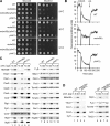
Mdm38 but not Ylh47 is required for respiratory growth. (A) Cells were cultured on YP glycerol medium to avoid the loss of mitochondrial DNA, washed, subjected to serial 10-fold dilutions, plated on YPD or YPG medium, and incubated at the indicated temperatures. (B) Δψ measurements by fluorescence quenching. (C) Mitochondrial proteins were subjected to SDS-PAGE and Western blotting. (D) Steady-state protein levels were analyzed as in C. Mitoch., mitochondria; prot., protein; val., valinomycin; WT, wild type.
Figure 3.
Figure 3.
Mdm38 is required for respiratory chain complex biogenesis. (A) Mitochondria were solubilized in 1% digitonin buffer, protein complexes were separated by BN-PAGE, and respiratory chain complexes were visualized by Western blot analysis. (B) After solubilization and BN-PAGE separation as in A, the F1Fo–ATPase complexes were analyzed by Western blot analysis. The asterisk indicates an unspecific protein band. M, monomer; WT, wild type.
Figure 4.
Figure 4.
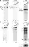
mdm38Δ mitochondria display defects in protein export. (A) In organello translation in the presence of [35S]methionine/cysteine (30 min) was performed in isolated wild-type (WT), mdm38Δ, and ylh47Δ mitochondria and unlabeled methionine was added and incubation continued for 5 min. Mitochondria were reisolated and subjected to SDS-PAGE and digital autoradiography. Atp9* indicates an SDS-resistant form of Atp9 seen in samples not TCA precipitated (Westermann et al., 2001). (B, top) In organello translation was performed for 15 min as in A followed by a 15-min chase. Mitochondria were subjected to osmotic shock, and samples were split and mock-treated or treated with 10 μg/ml proteinase K (Prot. K) for 15 min on ice. After reisolation and TCA precipitation, samples were subjected to SDS-PAGE, Western blotting, or digital autoradiography. (bottom) Quantification of the protease-inaccessible amount of newly synthesized mitochondrial proteins as a percentage of the synthesized protein. SEM was calculated from three independent experiments. (C) In organello translations were performed as described in A and analyzed by SDS-PAGE and digital autoradiography. p, precursor; m, mature.
Figure 5.
Figure 5.
Mdm38 interacts with mitochondrially encoded proteins. (A) In organello translations were performed with wild-type (WT) and Mdm38 Protein A–tagged mitochondria (Mdm38ProtA). After translation, mitochondria were lysed in digitonin buffer and subjected to IgG chromatography. Samples were eluted and subjected to SDS-PAGE and digital autoradiography or Western blotting. 10% of total and 100% of eluate were loaded. Atp9* indicates SDS-resistant Atp9. (B) 35S-labeled Qcr8 and Atp16 were imported into Tim18ProtA and Mdm38ProtA mitochondria. Mitochondria were treated with proteinase K, solubilized in digitonin buffer, and subjected to IgG chromatography. Bound proteins were analyzed as in A. 10% of total and 100% of eluate were loaded. White lines indicate that intervening lanes have been spliced out.
Figure 6.
Figure 6.
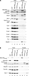
Protein interactions of Mdm38 and Ylh47. (A) Wild-type (WT), Mdm38ProtA, and Ylh47ProtA mitochondria were solubilized in digitonin buffer and subjected to IgG chromatography. Bound proteins were eluted with SDS sample buffer and analyzed by SDS-PAGE and Western blotting. 7% of total and 100% of eluate were loaded. (B) Mitochondria containing Mdm38ProtA, Oxa1ProtA, or Tim23ProtA were solubilized in digitonin buffer as described in A. The asterisk indicates the breakdown product of Oxa1ProtA. 7% of total and 100% of eluate were loaded. White lines indicate that intervening lanes have been spliced out.
Figure 7.
Figure 7.
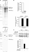
Mdm38 and Ylh47 form a complex with mitochondrial ribosomes. (A) Mitochondria were solubilized in digitonin buffer and subjected to IgG chromatography. The eluate was separated on urea SDS-PAGE and stained with colloidal Coomassie or analyzed by Western blotting with a Protein A antiserum. Lanes of the Coomassie-stained SDS gel were cut into slices and analyzed by mass spectrometry (Table I). *Mdm38ProtA indicates the breakdown product of Mdm38ProtA. (B) In organello translation was performed in isolated Mdm38ProtA mitochondria in the presence or absence of puromycin for 30 min. Mdm38ProtA was isolated as described in A. Samples were analyzed by SDS-PAGE, digital autoradiography, or Western blotting. n = 1. (C) After in organello translation, mitochondria were left untreated (lanes 1 and 3) or treated with puromycin (lane 2). After solubilization in low-salt (lanes 1 and 2) or high-salt (lanes 3) buffer, extracts were subjected to IgG chromatography and analyzed by SDS-PAGE and digital autoradiography or Western blotting. n = 1. 10% of total and 100% of eluate were loaded. White lines indicate that intervening lanes have been spliced out.

References

    1. Bonnefoy, N., M. Kermorgant, O. Groudinsky, M. Minet, P.P. Slonimski, and G. Dujardin. 1994. Cloning of a human gene involved in cytochrome oxidase assembly by functional complementation of an oxa1 − mutation in Saccharomyces cerevisiae. Proc. Natl. Acad. Sci. USA. 91:11978–11982. - PMC - PubMed
    1. Broadley, S.A., C.M. Demlow, and T.D. Fox. 2001. Peripheral mitochondrial inner membrane protein, Mss2p, required for export of the mitochondrially coded Cox2p C tail in Saccharomyces cerevisiae. Mol. Cell. Biol. 21:7663–7672. - PMC - PubMed
    1. Chacinska, A., M. Lind, A.E. Frazier, J. Dudek, C. Meisinger, A. Geissler, A. Sickmann, H.E. Meyer, K.N. Truscott, B. Guiard, et al. 2005. Mitochondrial presequence translocase: switching between TOM tethering and motor recruitment involves Tim21 and Tim17. Cell. 120:817–829. - PubMed
    1. DiMauro, S., and E.A. Schon. 2003. Mitochondrial respiratory-chain diseases. N. Engl. J. Med. 348:2656–2668. - PubMed
    1. Dimmer, K.S., S. Fritz, F. Fuchs, M. Messerschmitt, N. Weinbach, W. Neupert, and B. Westermann. 2002. Genetic basis of mitochondrial function and morphology in Saccharomyces cerevisiae. Mol. Biol. Cell. 13:847–853. - PMC - PubMed

Publication types

MeSH terms