. 2006 Feb 13;172(4):577-88.
doi: 10.1083/jcb.200510044.

A repeated IMP-binding motif controls oskar mRNA translation and anchoring independently of Drosophila melanogaster IMP

Affiliations

A repeated IMP-binding motif controls oskar mRNA translation and anchoring independently of Drosophila melanogaster IMP

Trent P Munro et al. J Cell Biol. .

Abstract

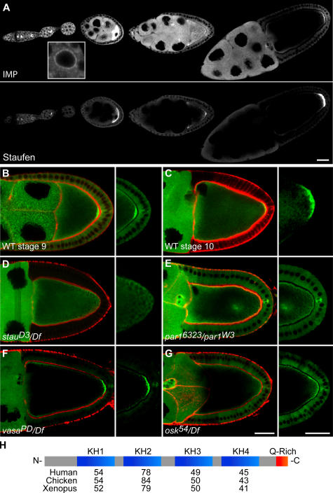
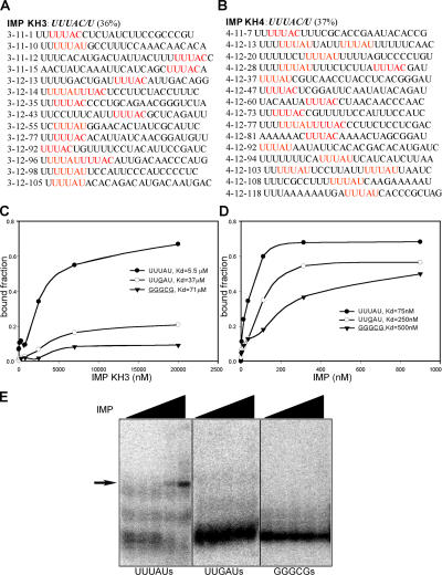
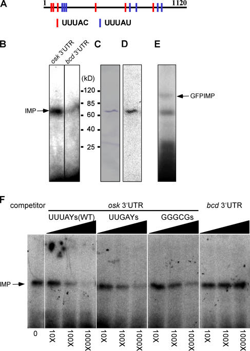
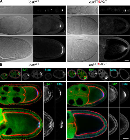
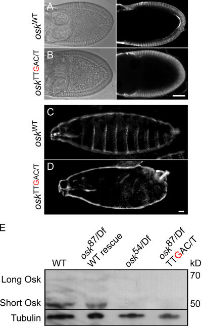
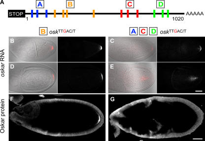
Zip code-binding protein 1 (ZBP-1) and its Xenopus laevis homologue, Vg1 RNA and endoplasmic reticulum-associated protein (VERA)/Vg1 RNA-binding protein (RBP), bind repeated motifs in the 3' untranslated regions (UTRs) of localized mRNAs. Although these motifs are required for RNA localization, the necessity of ZBP-1/VERA remains unresolved. We address the role of ZBP-1/VERA through analysis of the Drosophila melanogaster homologue insulin growth factor II mRNA-binding protein (IMP). Using systematic evolution of ligands by exponential enrichment, we identified the IMP-binding element (IBE) UUUAY, a motif that occurs 13 times in the oskar 3'UTR. IMP colocalizes with oskar mRNA at the oocyte posterior, and this depends on the IBEs. Furthermore, mutation of all, or subsets of, the IBEs prevents oskar mRNA translation and anchoring at the posterior. However, oocytes lacking IMP localize and translate oskar mRNA normally, illustrating that one cannot necessarily infer the function of an RBP from mutations in its binding sites. Thus, the translational activation of oskar mRNA must depend on the binding of another factor to the IBEs, and IMP may serve a different purpose, such as masking IBEs in RNAs where they occur by chance. Our findings establish a parallel requirement for IBEs in the regulation of localized maternal mRNAs in D. melanogaster and X. laevis.


Figures

Figure 1.
Figure 1.
IMP localization during oogenesis. (A–G) Localization of IMP during oogenesis visualized using G080, a GFP protein trap line (A, B, and E–G), or a GFP–IMP fusion construct specifically expressed in the germline (C and D). (A) IMP localizes to future oocyte in the germarium, accumulates in the oocyte through stage 7–8, appears enriched at both the anterior and posterior of the oocyte at stage 8, and is restricted to a crescent at the oocyte posterior pole by stage 9–10. These localizations are similar to those of Staufen protein, which marks the localization of osk mRNA. Within nurse cells, IMP is primarily cytoplasmic, but also rims the nucleus (inset). (B–G) IMP localization (green) and actin (red) in oocytes at stage 9 (B) or 10 (C) in WT and mutant backgrounds (D–G). (D) IMP is absent from the posterior crescent in a stau-null mutant that fails to localize osk RNA. (E) Like osk RNA (not depicted), IMP localizes ectopically in a transheterozygous par-1 allele combination that disrupts the polarity of the egg chamber. (F) IMP localizes normally in a vasa mutant that interferes with pole plasm formation. (G) IMP localizes to the posterior at stage 9 in an osk nonsense mutant (osk 54/Df(3R)pXT103) egg chamber, indicating its localization depends on osk RNA, not protein. (H) IMP contains four KH-type RNA-binding domains and a glutamine-rich COOH terminus. Numbers indicate the percentage of amino acid identity between IMP's KH domains and those of its homologues. Bars, 25 μm.
Figure 2.
Figure 2.
Characterization of the IBE UUUAY. (A and B) Representative RNA sequences selected after 12 rounds of SELEX against IMP's KH3 (A) and KH4 (B) domains. IBEs are in red. (C and D) Filter-binding assays between three tandem repeats of the winner sequence 4-12-13 (or the same RNA with IBEs mutated to UUgAU or gggcg) and either IMP KH3 (C) or the entire protein (D). (E) Electrophoretic mobility shift assay between IMP and three tandem copies of the winner sequence 4-12-13 containing either WT or mutant IBE motifs. Only RNAs with the WT (UUUAU) motifs induced a band shift.
Figure 3.
Figure 3.
The osk 3′UTR contains 13 IBEs and UV cross-links to IMP. (A) Positions of the 13 IBEs in the osk 3′UTR. (B) A 65-kD protein (IMP) in D. melanogaster ovary extracts specifically cross-links the 1,120-nt osk 3′UTR, but not the 817-nt bcd 3′UTR, which contains only two UUUAY motifs. (C and D) Anti-IMP immunoblot of ovary extract (C) labels the same band that UV cross-links to the 32P-osk 3′UTR (D). (E) 32P-osk 3′UTR cross-links to an ∼95-kD polypeptide (GFP–IMP) in embryo extracts of the protein trap line G080. (F) Cross-linking reactions between the 32P-osk 3′UTR and oocyte extracts in the presence of increasing concentrations of cold, competitor RNAs, including WT and mutant osk 3′UTRs and the bicoid 3′UTR.
Figure 4.
Figure 4.
osk mRNA, IMP, and Stau distributions in osk13 TTgAY flies. (A) Fluorescent in situ hybridizations comparing WT and mutant transgenic osk RNAs in flies that otherwise lack endogenous osk RNA. Mutant osk RNA localizes normally through stage 9 (middle), but by stage 10 (bottom), is evident as diffuse fluorescence fanning out from the posterior pole. (B) IMP (green) and Stau (blue) proteins in WT and mutant osk oocytes before stage 9 (top), at stage 9 (middle), and at stage 10 (bottom). Actin is visualized with rhodamine-phalloidin (red). At stage 9, Stau is localized normally at the posterior pole in oocytes that express the mutant osk transgene, whereas IMP is diffuse and not concentrated at the posterior. Both Stau and IMP are missing from the posterior pole in stage 10 oocytes that express the mutant osk RNA. Bars, 25 μm.
Figure 5.
Figure 5.
IBE mutations abolish osk RNA translational activation. (A and B) Anti-Osk immunostaining of osk 87/Df(3R)pXT103 egg chambers expressing a WT osk transgene, showing a crescent of Osk protein (A). Osk protein is missing in egg chambers from osk TT g AY flies (B). (C and D) Cuticle preparations of larvae from osk 87/Df(3R)pXT103 that express the WT (C) or mutant osk TTgAY (D) transgene. (E) Western blot of ovarian protein extracts probed with anti-Osk antibody, followed by an anti-tubulin antibody. Extracts were from WT flies with no osk transgene (WT) or from osk 87/Df(3R)pXT103 flies expressing either a WT or the IBE mutant osk TTgAY osk transgene, or the nonsense mutant osk 54 transgene. Neither long nor short Osk is present in the osk TTgAY or osk 54 mutants. Bars, 25 μm.
Figure 6.
Figure 6.
Mutations to nonoverlapping subsets of osk's IBEs and their affects on osk RNA and protein distributions in the oocyte. (A) Four subsets of IBEs in the osk 3′UTR: A, B, C, and D. (B–E) osk in situ hybridizations at stages 9 (B and C) and 10 (D and E). (F and G) Osk protein detected by immunofluorescence at stage 10. Oocytes that express osk RNA with mutations to IBE subset B display normal localization of osk mRNA and protein. Mutations to subset D, or to subsets A and C (not depicted) cause osk RNA delocalization from the posterior pole at stage 10; Osk protein is absent in these oocytes. Bars, 25 μm.
Figure 7.
Figure 7.
Analysis of oocytes that lack IMP protein. (A) The imp gene is flanked by sesB, Ant2, and sbr. Diagram shows alternatively spliced isoforms (exons in blue), the location of GFP in protein trap line G080, and P element insertion EP(X)760 used to create three mutant alleles of IMP. The positions of the two alternate ATGs are shown in green. (B) Immunoblots of preblastoderm embryos laid by mothers with IMP mutant germline clones. IMP2 may produce a truncated protein, whereas IMP7 and IMP8 are protein nulls. (C, top) A germline clone (center egg chamber) marked by the absence of GFP. (bottom) IMP is absent only from the mutant clone. (D) The distributions of osk mRNA and Osk protein in IMP-null egg chambers (middle and right) are indistinguishable from WT (left) in IMP mutant germline clones generated using the FLP/FRT OvoD1 DFS technique. Bars, 25 μm.

References

    1. Bergsten, S.E., and E.R. Gavis. 1999. Role for mRNA localization in translational activation but not spatial restriction of nanos RNA. Development. 126:659–669. - PubMed
    1. Bohl, F., C. Kruse, A. Frank, D. Ferring, and R.P. Jansen. 2000. She2p, a novel RNA-binding protein tethers ASH1 mRNA to the Myo4p myosin motor via She3p. EMBO J. 19:5514–5524. - PMC - PubMed
    1. Brand, A.H., and N. Perrimon. 1993. Targeted gene expression as a means of altering cell fates and generating dominant phenotypes. Development. 118:401–415. - PubMed
    1. Breitwieser, W., F.H. Markussen, H. Horstmann, and A. Ephrussi. 1996. Oskar protein interaction with Vasa represents an essential step in polar granule assembly. Genes Dev. 10:2179–2188. - PubMed
    1. Bubunenko, M., T.L. Kress, U.D. Vempati, K.L. Mowry, and M.L. King. 2002. A consensus RNA signal that directs germ layer determinants to the vegetal cortex of Xenopus oocytes. Dev. Biol. 248:82–92. - PubMed

Publication types