. 2006 Feb 13;172(4):619-31.
doi: 10.1083/jcb.200505138.

The tumor suppressor DAPK inhibits cell motility by blocking the integrin-mediated polarity pathway

Affiliations

The tumor suppressor DAPK inhibits cell motility by blocking the integrin-mediated polarity pathway

Jean-Cheng Kuo et al. J Cell Biol. .

Abstract

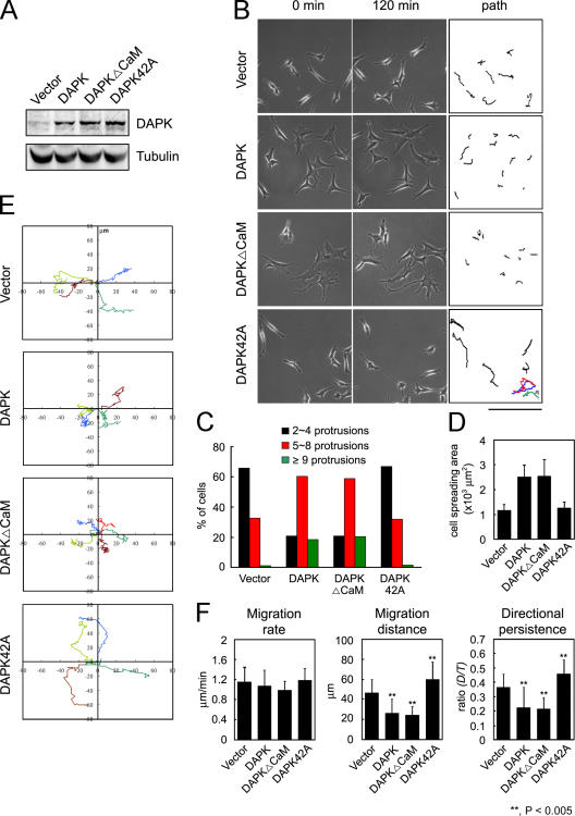
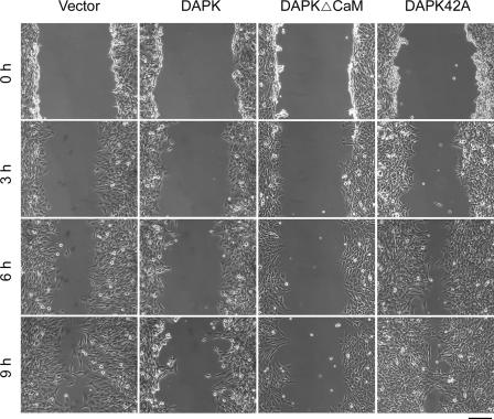
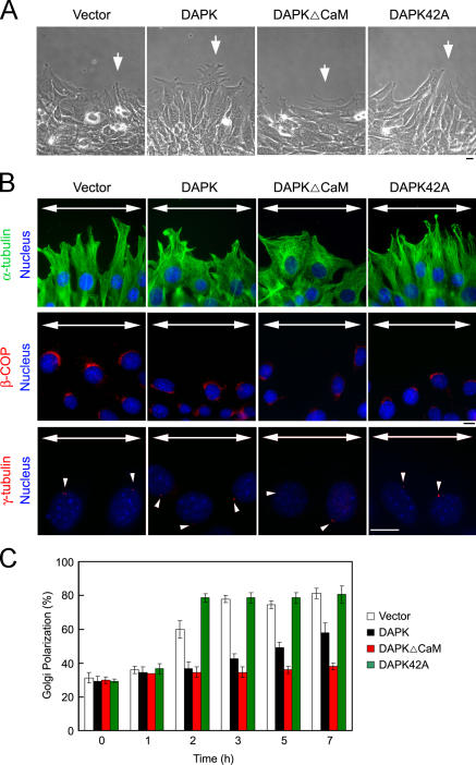
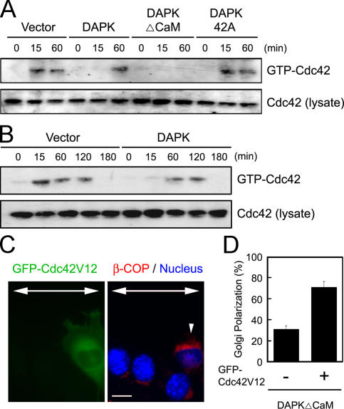
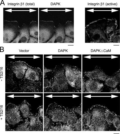
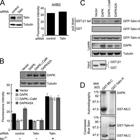
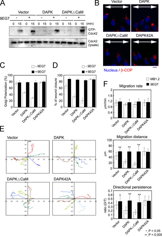
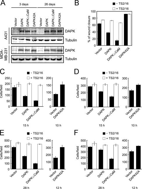
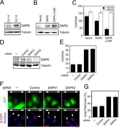
Death-associated protein kinase (DAPK) is a calmodulin-regulated serine/threonine kinase and possesses apoptotic and tumor-suppressive functions. However, it is unclear whether DAPK elicits apoptosis-independent activity to suppress tumor progression. We show that DAPK inhibits random migration by reducing directional persistence and directed migration by blocking cell polarization. These effects are mainly mediated by an inhibitory role of DAPK in talin head domain association with integrin, thereby suppressing the integrin-Cdc42 polarity pathway. We present evidence indicating that the antimigratory effect of DAPK represents a mechanism through which DAPK suppresses tumors. First, DAPK can block migration and invasion in certain tumor cells that are resistant to DAPK-induced apoptosis. Second, using an adenocarcinoma cell line and its highly invasive derivative, we demonstrate DAPK level as a determining factor in tumor invasiveness. Collectively, our study identifies a novel function of DAPK in regulating cell polarity during migration, which may act together with its apoptotic function to suppress tumor progression.


Figures

Figure 1.
Figure 1.
Effects of DAPK on random migration. (A) NIH3T3 cells were infected with recombinant retrovirus carrying vector or various forms of DAPK, selected by puromycin, and subjected to immunoblot analyses with antibodies as indicated. (B) Morphologies and migratory behaviors of NIH3T3 cells expressing various DAPK proteins. Cells as in A were plated and monitored 30 h after plating. (left and middle) Images at 0 and 120 min, respectively. (right) These images delineate the path of each cell during the 120-min period. Colored lines are used to distinguish overlapped cell paths. Video 1 corresponds to this figure. (C) Regulation of cell protrusions by DAPK. Protrusions longer than 5 μm formed per cell were quantitated based on time-lapse images. The percentage of cells in each population having protrusions within the indicated ranges was calculated and plotted. For each cell population, at least 300 cells were scored. (D) Areas of spreading were measured using Metamorph software and plotted. Data shown are means ± SD. n = 50. (E) Analysis of migration paths. The tracks of representative cells shown in B were plotted. The origins of migration were superimposed at 0, 0. (F) Migration parameters were calculated as described in Materials and methods. Data represent means ± SD from trajectories of 30 cells. **, P < 0.005, as compared with cells with control vector. Video 1 is available at http://www.jcb.org/cgi/content/full/jcb.200505138/DC1. Bar, 200 μm.
Figure 2.
Figure 2.
Effect of DAPK on wound-healing migration. Cells, as in Fig. 1, were assayed for wound-healing migration, and cell migration into wounds was monitored by time-lapse microscopy. Still images were captured at the indicated times after wounding. Bar, 200 μm.
Figure 3.
Figure 3.
DAPK interferes with cell polarization during wound-healing migration. (A) The morphologies of wound-edge cells. A confluent monolayer of cells, as in Fig. 2, was wounded, and still images were taken 5 h after wounding. Arrows indicate the morphological differences of each population of cells at the wound edge. Videos 2–5 correspond to these images. (B) The effects of DAPK on microtubule polarization and Golgi and MTOC reorientation. Cells as in A were wounded and fixed at 5 h after wounding. Cells were double stained with Hoechst 33342 and anti–α-tubulin (to visualize microtubules), anti–β-COP (to visualize Golgi), or anti–γ-tubulin antibody (to visualize MTOC) and examined by fluorescence microscopy. Arrows indicate the direction of the wound, and arrowheads mark the locations of MTOC. (C) Kinetics of Golgi reorientation. The percentage of wound-edge cells with their Golgi apparatus in the forward-facing 120° sector was measured at the indicated time points after wounding. For each time point and cell population, at least 150 cells were scored. Data represent means ± SD. n = 3. Videos 2–5 are available at http://ww.jcb.org/cgi/content/full/jcb.200505138/DC1. Bars, 10 μm.
Figure 4.
Figure 4.
DAPK disrupts cell polarity by blocking Cdc42 activation. (A and B) NIH3T3 cells expressing various DAPK proteins were wounded as described in Materials and methods and lysed at indicated time points after wounding. (top) The amount of GTP-bound Cdc42 was determined by GST-PAK-CRIB pull-down analysis, followed by Western blotting with anti-Cdc42 antibody. (bottom) The levels of total Cdc42. (C) NIH3T3 cells expressing DAPKΔCaM were transiently transfected with pGFP-C1-Cdc42V12 and assayed for wound-healing migration. Cells were fixed 5 h after wounding and stained with anti–β-COP antibody and Hoechst 33342. Arrows indicate the direction of the wound, and the arrowhead marks the GFP-positive cell (right). (D) The percentage of GFP-positive and -negative cells, as in C, with polarized Golgi was measured in the front row cells. For each population, at least 150 cells were scored. Data represent means ± SD. n = 3. Bar, 10 μm.
Figure 5.
Figure 5.
Both wound-induced activation of integrin β1 and DAPK-induced blockage of integrin signaling occur at the leading edge. (A) Activation of integrin β1 at the leading edge of a migrating cell. A monolayer of MDA-MB-231 cells was scratched and fixed 5 h later. Cells were double stained for total integrin β1 (with antibody P4C10) and DAPK or stained for active integrin β1 (with antibody B44) and examined by confocal microscopy. (B) DAPK blocks wound-induced FAK activation at the leading edge. MDA-MB-231 cells stably expressing various DAPK proteins (Fig. 8 A) were treated with or without TS2/16 before wounding and fixed 5 h later. Cells were stained with p-FAK and examined by confocal microcopy. Arrows indicate the direction of the wound. Bars, 10 μm.
Figure 6.
Figure 6.
DAPK interferes with talin-H binding to integrin. (A) 293T cells were transfected with talin or control siRNA as indicated. Transfection efficiency was >90%. The expression of talin and tubulin were detected by Western blot (left), and the surface expression of integrin β1 was determined by flow cytometry analysis with antibody AIIB2 (right). (B) 293T cells cotransfected with talin or control siRNA and various DAPK constructs were treated with or without Mn2+ and then analyzed for cell surface binding of B44 (bottom). (top) The expression of various DAPK proteins. For the flow cytometry analyses in A and B, data shown are mean fluorescence intensities of B44 or AIIB2 binding subtracted by the background fluorescence intensities obtained from experiments using only the secondary antibody. n = 3. (C) 293T cells transfected with various forms of DAPK and GFP–talin-H were cultured in suspension and then lysed. Cell lysates were incubated with GST or GST-β1 tail, and GFP–talin-H bound on beads was analyzed by Western blot (top). The expression of various proteins in cell lysates was analyzed by Western blot (middle). (bottom) The equal input of GST and GST-β1 tail. (D) DAPK does not phosphorylate talin-H. Flag-tagged DAPK immunoprecipitated from lysate of transfected cells was used to phosphorylate bacterially expressed GST−myosin light chain (MLC) or GST–talin-H. Phosphorylated proteins were detected by autoradiography, and the positions of GST-MLC and autophosphorylated DAPK are indicated (top). The equal input of GST fusion proteins (*) is shown on the bottom.
Figure 7.
Figure 7.
Activation of integrin β1 rescues DAPK-induced migratory defects. (A) A monolayer of NIH3T3 cells expressing various DAPK proteins was treated with or without 9EG7 before scratch. Cells were harvested 0 or 15 min after scratch and the amounts of GTP-bound and total Cdc42 were determined. (B) Cells as in A were analyzed for Golgi polarization 5 h after wounding. Arrows indicate the direction of the wound. (C) The percentage of cells, as in B, with polarized Golgi was quantitated. (D) A monolayer of NIH3T3 derivatives was treated with or without 9EG7 and assayed for wound-healing migration. The percentage of wound closure at 9 h after wounding was calculated. Data represent means ± SD. n = 3. (E) NIH3T3 cells expressing various DAPK proteins were treated with or without 9EG7 and assayed for random migration. The paths of representative cells during a 120-min period were plotted. (F) Cells, as in E, were assayed for free migration in the presence of 5 μg/ml 9EG7 or MB1.2 antibody. Migration parameters were calculated as in Fig. 1. Data represent means ± SD from trajectories of 30 cells. *, P < 0.05; **, P < 0.005, as compared with cells treated with MB1.2. Bar, 10 μm.
Figure 8.
Figure 8.
DAPK inhibits migration and invasion in p53-defective tumor cell lines. (A) A431 and MDA-MB-231 cells were infected with retrovirus-carrying vector or various forms of DAPK, selected by puromycin, left in culture for 3 or 26 d, and subjected to immunoblot analysis to detect the expression of DAPK proteins. (B) A431 cells were infected as in A, selected by puromycin for 3 d, and assayed for wound-healing migration in the presence or absence of TS2/16. The percentage of wound closure at 8 h after wounding was calculated. (C and D) The effect of DAPK on haptotactic migration. A431 (C) or MDA-MB-231 (D) cells expressing various DAPK proteins were assayed for haptotactic migration in the presence or absence of TS2/16. After the indicated time periods, cells that migrated to the lower side of the chamber were counted. (E and F) The effect of DAPK on invasion. A431 (E) or MDA-MB-231 (F) cells stably expressing various DAPK proteins were assayed for invasion through Matrigel. After the indicated time periods, cells invaded through Matrigel were counted. Data represent means ± SD. n = 3.
Figure 9.
Figure 9.
DAPK level is a determining factor in tumor invasion. (A) Analysis of DAPK expression in CL1-0 and CL1-5 cells by immunoblot analysis. (B) CL1-5 cells were transfected with DAPK or DAPKΔCaM and subjected to immunoblot analysis to detect the expression of DAPK proteins. (C) CL1-5 cells were transiently cotransfected with vector or various forms of DAPK, together with GFP, at a ratio of 4:1. Cells were treated with or without TS2/16 and assayed for their invasiveness. After 20 h of incubation, the GFP-positive cells invaded through Matrigel were counted. Data represent means ± SD. (D) CL1-0 cells were transiently transfected with control siRNA, DAPK siRNA1, or DAPK siRNA2, together with an expression vector for CD2-FAK. Transfected cells were isolated by the CELLetion CD2 kit, lysed, and subjected to immunoblot analysis to detect the expression of DAPK proteins. (E) CL1-0 cells were transfected with various siRNAs together with GFP at a ratio of 9:1. Transfected cells were assayed for their invasiveness, as in C, and the GFP-positive cells invaded through Matrigel were counted at 48 h after incubation. (F) A monolayer of CL1-0 cells transfected as in E was wounded. Cells were fixed 5 h after wounding and stained with anti–β-COP and Hoechst 33342. Arrows indicate the direction of the wound and small arrowheads mark the GFP-positive cells at the wound edge. (G) The percentage of GFP-positive cells as in F with polarized Golgi was measured in the front row cells. For each cell population, at least 150 cells were scored. Data represent means ± SD. n = 3. Bar, 10 μm.

References

    1. Allen, W.E., D. Zicha, A.J. Ridley, and G.E. Jones. 1998. A role for Cdc42 in macrophage chemotaxis. J. Cell Biol. 141:1147–1157. - PMC - PubMed
    1. Bialik, S., and A. Kimchi. 2004. DAP-kinase as a target for drug design in cancer and diseases associated with accelerated cell death. Semin. Cancer Biol. 14:283–294. - PubMed
    1. Bialik, S., A.R. Bresnick, and A. Kimchi. 2004. DAP-kinase-mediated morphological changes are localization dependent and involve myosin-II phosphorylation. Cell Death Differ. 11:631–644. - PubMed
    1. Calderwood, D.A., B. Yan, J.M. de Pereda, B.G. Alvarez, Y. Fujioka, R.C. Liddington, and M.H. Ginsberg. 2002. The phosphotyrosine binding-like domain of talin activates integrins. J. Biol. Chem. 277:21749–21758. - PubMed
    1. Calderwood, D.A., Y. Fujioka, J.M. de Pereda, B. Garcia-Alvarez, T. Nakamoto, B. Margolis, C.J. McGlade, R.C. Liddington, and M.H. Ginsberg. 2003. Integrin beta cytoplasmic domain interactions with phosphotyrosine-binding domains: a structural prototype for diversity in integrin signaling. Proc. Natl. Acad. Sci. USA. 100:2272–2277. - PMC - PubMed

Publication types

MeSH terms