. 2006 Feb 21;113(7):1005-14.
doi: 10.1161/CIRCULATIONAHA.105.588954. Epub 2006 Feb 13.

In vivo visualization of embryonic stem cell survival, proliferation, and migration after cardiac delivery

Affiliations

In vivo visualization of embryonic stem cell survival, proliferation, and migration after cardiac delivery

Feng Cao et al. Circulation. .

Abstract

Background: Recent studies have shown that stem cell therapy can promote tissue regeneration; however, monitoring stem cells in vivo remains problematic owing to limitations of conventional histological assays and imaging modalities.

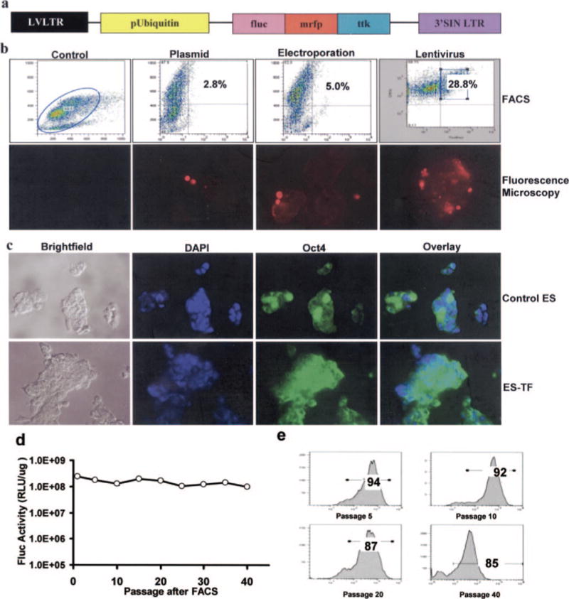
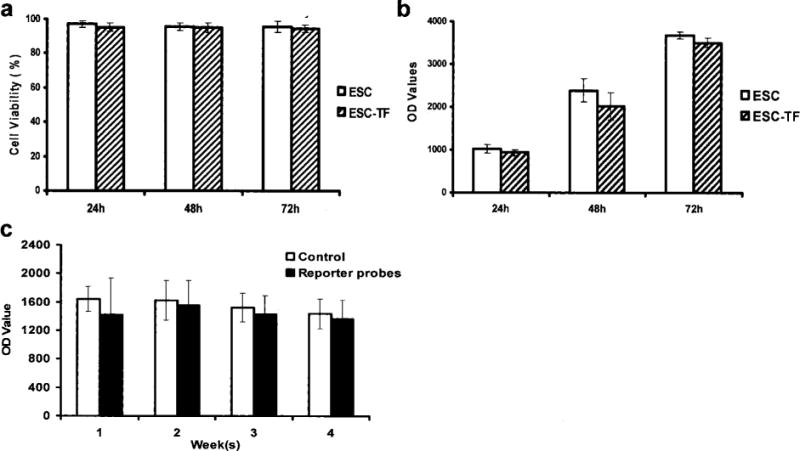
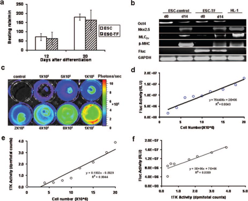
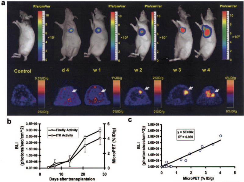
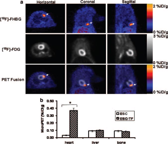
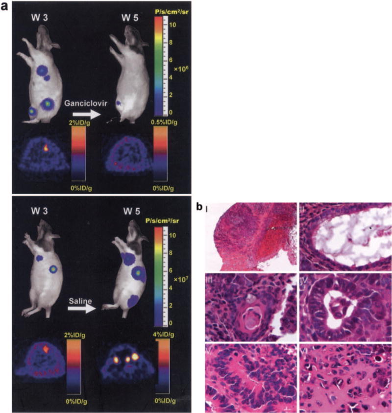
Methods and results: Murine embryonic stem (ES) cells were stably transduced with a lentiviral vector carrying a novel triple-fusion (TF) reporter gene that consists of firefly luciferase, monomeric red fluorescence protein, and truncated thymidine kinase (fluc-mrfp-ttk). ES cell viability, proliferation, and differentiation ability were not adversely affected by either reporter genes or reporter probes compared with nontransduced control cells (P=NS). Afterward, 1x10(7) of ES cells carrying the TF reporter gene (ES-TF) were injected into the myocardium of adult nude rats (n=20). Control animals received nontransduced ES cells (n=6). At day 4, the bioluminescence and positron emission tomography signals in study animals were 3.7x10(7)+/-5.8x10(6) photons.s(-1).cm(-2) per steradian (sr) and 0.08+/-0.03% injected dose/g, respectively (P<0.05 versus control). Both signals increased progressively from week 1 to week 4, which indicated ES cell survival and proliferation in the host. Histological analysis demonstrated the formation of intracardiac and extracardiac teratomas. Finally, animals (n=4) that were treated with intraperitoneal injection of ganciclovir (50 mg/kg) did not develop teratomas when compared with control animals (n=4) treated with saline (1 mL/kg).

Conclusions: This is the first study to characterize ES cells that stably express fluorescence, bioluminescence, and positron emission tomography reporter genes and monitor the kinetics of ES cell survival, proliferation, and migration. This versatile imaging platform should have broad applications for basic research and clinical studies on stem cell therapy.


Figures

Figure 1
Figure 1
Stable lentiviral transduction of ES cells with the TF reporter gene. a, Schema of the TF reporter gene containing fusion of fluc-mrfp-ttk. The TF reporter gene was cloned into a self-inactivating lentiviral vector downstream from the ubiquitin promoter. The 3 fusion proteins are joined by a 14-amino acid (LENSHASAGYQAST) and 8-amino acid (TAGPGSAT) linker, respectively. b, FACS histograms of ES cells 48 hours after transduction with plasmid lipofectamine, electroporation, and lentivirus carrying the TF reporter gene. c, Control ES and ES-TF cells showed similar morphology and stem cell marker Oct4 expression on brightfield and fluorescence microscopy, respectively. DAPI staining is used as a nuclear marker. The stability of TF reporter gene expression was measured quantitatively by both (d) fluc enzyme assays and (e) mrfp FACS for 40 passages. LVLTR indicates lentivirus long-terminal repeat.
Figure 2
Figure 2
Effects of reporter genes and reporter probes on cell viability, proliferation, and differentiation. Trypan blue cell viability (a) and CyQuant cell proliferation assay (b) both showed no significant difference between control ES and ES-TF cells at various time points. c, Weekly exposure of ES-TF cells to combined bioluminescence reporter probe D-luciferin (100 μmol/L) and PET reporter probe [18F]-FHBG (100 μCi) had no significant adverse effects on ES-TF cell proliferation in vitro over 1 month.
Figure 3
Figure 3
Effect of TF reporter gene expression on ES cell differentiation. a, Both control ES and ES-TF cells showed similar beating rates per minute at day 12 and day 20 of embryoid body differentiation (P<0.05 vs day 12). b, RT-PCR analysis showed the levels of cardiac transcriptional factor (Nkx2.5) and ventricular-specific marker (β-MHC, MLC2v) increased from day 0 to day 14 of embryoid body differentiation, whereas the stem cell markers (Oct4) decreased during the same period. Fluc is present only within ESC-TF cells, as expected. HL-1 is a control mouse cardiomyocyte cell line that is positive for Nkx2.5 and β-MHC but negative for Oct4. GAPDH is a loading control for all cells. c, Bioluminescence imaging of varying numbers of ES-TF cells plated on 24-well plates with increasing signals. P/sec indicates photons · sec−1 · cm−2 · sr−1. Stably transduced ES-TF cells showed robust correlation with (d) fluc enzyme activity (r2=0.93) and (e) ttk enzyme activity (r2=0.90). f, Fluc and ttk activities also correlated well with each other (r2=0.83). RLU indicates relative light units; dpm, disintegrations per minute.
Figure 4
Figure 4
Molecular imaging of transplanted ES cells with bioluminescence and PET imaging. a, To assess longitudinal cell survival, animals were imaged for 4 weeks. A representative study animal injected with ES-TF cells showed significant bioluminescence (top) and PET (bottom) signals at day 4, week 1, week 2, week 3, and week 4. In contrast, control animals had background activities only. b, Quantification of imaging signals showed a drastic increase of fluc and ttk activities from week 2 to week 4. Extracardiac signals were observed during subsequent weeks. c, Quantification of cell signals showed a robust in vivo correlation between bioluminescence and PET imaging (r2=0.92). BLI indicates bioluminescence.
Figure 5
Figure 5
Defining the tomographic location of transplanted ES cells in the myocardium. a, Two weeks after cell transplantation, animals underwent [18F]-FHBG reporter probe imaging (top row) followed by [18F]-FDG myocardial viability imaging (middle row). Fusion of [18F]-FHBG and [18F]-FDG images (bottom row) shows the exact anatomic location of transplanted ES-TF cells (arrow) at the anterolateral wall in horizontal, coronal, and sagittal views. b, The cardiac [18F]-FHBG activities between control ES and ES-TF animals were 0.03±0.01% ID/g and 0.37±0.05% ID/g, respectively (*P<0.05). Notice that for the [18F]-FHBG image, there is residual activity in the liver and bone due to hepatic clearance of tracer and free fluoride in systemic circulation.
Figure 6
Figure 6
Ablation of teratoma formation with ttk as both a reporter gene and a suicide gene. a, Treatment of control animals with saline resulted in multiple teratoma formation by week 5. In contrast, study animals treated with ganciclovir for 2 weeks showed abrogation of both bioluminescence and PET imaging signals. b, Postmortem histological analysis of a representative explanted heart showed (I) anterior wall of the myocardium injected with cells surrounding area of hemorrhage (40×); (II) respiratory epithelium with ciliated columnar and mucin-producing goblet cells (1000×); (III) squamous cell differentiation with keratin pearl; (IV) nonciliated columnar gland (1000×); (V) rosette consistent with neuroectodermal differentiation (1000×); and (VI) osteoid (nonmineralized bone) formation (1000× magnification).

References

    1. Eyre H, Kahn R, Robertson RM, Clark NG, Doyle C, Hong Y, Gansler T, Glynn T, Smith RA, Taubert K, Thun MJ. Preventing cancer, cardiovascular disease, and diabetes: a common agenda for the American Cancer Society, the American Diabetes Association, and the American Heart Association. Circulation. 2004;109:3244–3255. - PubMed
    1. Orlic D, Kajstura J, Chimenti S, Jakoniuk I, Anderson SM, Li B, Pickel J, McKay R, Nadal-Ginard B, Bodine DM, Leri A, Anversa P. Bone marrow cells regenerate infarcted myocardium. Nature. 2001;410:701–705. - PubMed
    1. Kocher AA, Schuster MD, Szabolcs MJ, Takuma S, Burkhoff D, Wang J, Homma S, Edwards NM, Itescu S. Neovascularization of ischemic myocardium by human bone-marrow-derived angioblasts prevents cardiomyocyte apoptosis, reduces remodeling and improves cardiac function. Nat Med. 2001;7:430–436. - PubMed
    1. Hodgson DM, Behfar A, Zingman LV, Kane GC, Perez-Terzic C, Alekseev AE, Puceat M, Terzic A. Stable benefit of embryonic stem cell therapy in myocardial infarction. Am J Physiol Heart Circ Physiol. 2004;287:H471–H479. - PubMed
    1. Wollert KC, Drexler H. Clinical applications of stem cells for the heart. Circ Res. 2005;96:151–163. - PubMed

Publication types

MeSH terms