

The structure of a ring-opened proliferating cell nuclear antigen-replication factor C complex revealed by fluorescence energy transfer
- PMID: 16476998
- PMCID: PMC1413853
- DOI: 10.1073/pnas.0511263103
The structure of a ring-opened proliferating cell nuclear antigen-replication factor C complex revealed by fluorescence energy transfer
Abstract
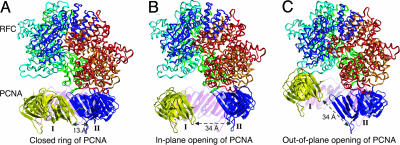
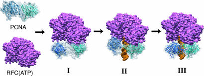
Numerous proteins that function in DNA metabolic pathways are known to interact with the proliferating cell nuclear antigen (PCNA). The important function of PCNA in stimulating various cellular activities requires its topological linkage with DNA. Loading of the circular PCNA onto duplex DNA requires the activity of a clamp-loader [replication factor C (RFC)] complex and the energy derived from ATP hydrolysis. The mechanistic and structural details regarding PCNA loading by the RFC complex are still developing. In particular, the positive identification of a long-hypothesized structure of an open clamp-RFC complex as an intermediate in loading has remained elusive. In this study, we capture an open yeast PCNA clamp in a complex with RFC through fluorescence energy transfer experiments. We also follow the topological transitions of PCNA in the various steps of the clamp-loading pathway through both steady-state and stopped-flow fluorescence studies. We find that ATP effectively drives the clamp-loading process to completion with the formation of the closed PCNA bound to DNA, whereas ATPgammaS cannot. The information derived from this work complements that obtained from previous structural and mechanistic studies and provides a more complete picture of a eukaryotic clamp-loading pathway using yeast as a paradigm.
Conflict of interest statement
Conflict of interest statement: No conflicts declared.
Figures
References
-
- Garg P., Burgers P. M. Crit. Rev. Biochem. Mol. Biol. 2005;40:115–128. - PubMed
-
- Kuriyan J., O'Donnell M. J. Mol. Biol. 1993;234:915–925. - PubMed
-
- Trakselis M. A., Benkovic S. J. Structure (Cambridge, U.K.) 2001;9:999–1004. - PubMed
-
- Prelich G., Tan C. K., Kostura M., Mathews M. B., So A. G., Downey K. M., Stillman B. Nature. 1987;326:517–520. - PubMed
-
- Krishna T. S., Kong X. P., Gary S., Burgers P. M., Kuriyan J. Cell. 1994;79:1233–1243. - PubMed
Publication types
MeSH terms
Substances
Grants and funding
LinkOut - more resources
Full Text Sources
Molecular Biology Databases
Miscellaneous
