Crystal structure of the IL-2 signaling complex: paradigm for a heterotrimeric cytokine receptor
- PMID: 16477002
- PMCID: PMC1413841
- DOI: 10.1073/pnas.0511161103
Crystal structure of the IL-2 signaling complex: paradigm for a heterotrimeric cytokine receptor
Abstract
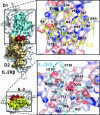
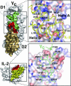
IL-2 is a cytokine that functions as a growth factor and central regulator in the immune system and mediates its effects through ligand-induced hetero-trimerization of the receptor subunits IL-2R alpha, IL-2R beta, and gamma(c). Here, we describe the crystal structure of the trimeric assembly of the human IL-2 receptor ectodomains in complex with IL-2 at 3.0 A resolution. The quaternary structure is consistent with a stepwise assembly from IL-2/IL-2R alpha to IL-2/IL-2R alpha/IL-2R beta to IL-2/IL-2R alpha/IL-2R beta/gamma(c). The IL-2R alpha subunit forms the largest of the three IL-2/IL-2R interfaces, which, together with the high abundance of charge-charge interactions, correlates well with the rapid association rate and high-affinity interaction of IL-2R alpha with IL-2 at the cell surface. Surprisingly, IL-2R alpha makes no contacts with IL-2R beta or gamma(c), and only minor changes are observed in the IL-2 structure in response to receptor binding. These findings support the principal role of IL-2R alpha to deliver IL-2 to the signaling complex and act as regulator of signal transduction. Cooperativity in assembly of the final quaternary complex is easily explained by the extraordinarily extensive set of interfaces found within the fully assembled IL-2 signaling complex, which nearly span the entire length of the IL-2R beta and gamma(c) subunits. Helix A of IL-2 wedges tightly between IL-2R beta and gamma(c) to form a three-way junction that coalesces into a composite binding site for the final gamma(c) recruitment. The IL-2/gamma(c) interface itself exhibits the smallest buried surface and the fewest hydrogen bonds in the complex, which is consistent with its promiscuous use in other cytokine receptor complexes.
Conflict of interest statement
Conflict of interest statement: No conflicts declared.
Figures


References
-
- Morgan D. A., Ruscetti F. W., Gallo R. Science. 1976;193:1007–1008. - PubMed
-
- Robb R. J., Smith K. A. Mol. Immunol. 1981;18:1087–1094. - PubMed
-
- Smith K. A., Favata M. F., Oroszlan S. J. Immunol. 1983;131:1808–1815. - PubMed
-
- Taniguchi T., Matsui H., Fujita T., Takaoka C., Kashima N., Yoshimoto R., Hamuro J. Nature. 1983;302:305–310. - PubMed
Publication types
MeSH terms
Substances
Associated data
- Actions
LinkOut - more resources
Full Text Sources
Other Literature Sources
Molecular Biology Databases

