. 2006 Feb 21;103(8):2752-7.
doi: 10.1073/pnas.0510970103. Epub 2006 Feb 13.

Negative regulation of activation-induced cytidine deaminase in B cells

Affiliations

Negative regulation of activation-induced cytidine deaminase in B cells

Taro Muto et al. Proc Natl Acad Sci U S A. .

Abstract

Both class switch recombination (CSR) and somatic hypermutation (SHM) of the Ig genes require the activity of activation-induced cytidine deaminase (AID). Expression of AID is restricted to B cells in the germinal centers of the lymphoid organs, where activated B cells undergo CSR and SHM. We previously showed that constitutive and systemic expression of AID leads to tumorigenesis in T cells and lung epithelium, but not in B cells. This finding led us to suspect that transgenic AID may be inactivated at least in part in B cells. To address this issue, we generated conditional AID-transgenic mice that constitutively express AID only in B cells. Studies on the cross between the AID-transgenic and AID-deficient mice showed that abundant AID protein accumulated by constitutive expression is inactivated in B cells, possibly providing an explanation for the absence of deregulation of CSR and SHM in AID-transgenic B cells.


Conflict of interest statement

Conflict of interest statement: No conflicts declared.

Figures
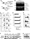
Fig. 1.
Fig. 1.
B cell-specific expression of AID in the double-Tg mice. (A) The conditional AID transgene construct that expresses AID after Cre-dependent recombination. The floxed GFP located upstream of AID cDNA is regulated under the transcriptional control of the constitutive and ubiquitous promoter pCAG. The position of primer pairs that detect both the intact and recombined forms of constructs are shown under the construct by arrowheads, and the sizes of amplified bands are shown. (B) PCR analysis of the recombination of the transgene DNA in various tissues of the Tg mice with or without CD19-cre. (C) Flow cytometric profiles of B lymphocytes from several lymphoid organs of the double-Tg mice after staining with antibodies for B cell markers (B220 or CD19). Numbers indicate percentages of B cell marker+ GFP cells in lymphocytes. (D) The percentage of the GFP cells in the B cell marker (B220 or CD19)-positive (B cell) and -negative cells (Non-B cell). Results obtained from three individual mice aged 8–9 weeks are summarized as column graphs with mean ± SD values. (E) GFP depletion in developing B cells of the double-Tg mice. Pro- and pre-B cells in BM are defined as c-kit+ B220+ and BP1+ B220+, respectively. Mature B cells in SPL are defined as IgM+ CD19+. (F) Northern blot analysis of endogenous and Tg AID expression in the lymphoid tissues of wild-type and Tg mice. Total RNA from unstimulated SPL, MLN, and PP were extracted and electrophoresed. Blots were probed for AID mRNA and rehybridized with a GAPDH probe after stripping the AID signal. RNA samples from stimulated and unstimulated CH12F3–2A (CH12) cells were prepared for endogenous AID control. RNAs from the HEK293T (293T) cells transfected with the transgene vector with or without Cre-expressing vector are used for controls of transgene AID before or after recombination, respectively. To compare the amounts of endogenous and Tg AID mRNA, RNA was prepared from purified GFP B220+ SPL B cells of the double-Tg mice after stimulation by LPS for 3 days (stimulated SPL cells). (G) AID protein production from the double-Tg mice with AID−/− background. AID protein in splenocytes of the double-Tg × AID−/− mice and their controls after stimulation with LPS for 4 days was immunoprecipitated by an anti-AID monoclonal antibody (clone 243) and subjected to immunoblot by the same antibody after SDS/PAGE. Threefold dilutions of input amounts are shown. Nonspecific signal (NS) from the blot is shown as an internal control to indicate input protein amounts.
Fig. 2.
Fig. 2.
Normal B cell development in BM and the secondary lymphoid organs in the double-Tg mice. (A) Flow cytometric analysis of B cells in SPL, MLN, and PP from the Tg mice. The percentages of PNA+ cells in the CD19+ population are indicated. Results obtained from three individual mice aged 8–9 weeks are summarized as column graphs with mean ± SD values. (B) Flow cytometric analysis of the BM cells from the Tg mice. The percentages of pro-B, pre-B, and immature B cells in the B220+ cell population in BM are indicated. Pro-B, c-kit+ B220+; pre-B, BP1+ B220+; immature B, B220lo IgM+. (C) The percentages of IgM+ and IgG+ cells in the CD19+ population are indicated. Results obtained from three individual mice aged 8–9 weeks are summarized as column graphs with mean ± SD values.
Fig. 3.
Fig. 3.
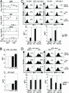
Analyses of CSR induced by Tg AID. (A) Serum Ig concentrations in the AID-Tg mice that lack endogenous AID and their control mice (aged 10–15 weeks) were determined by ELISA. (B) In vitro class switching of splenocytes from the AID-Tg mice in the presence of endogenous AID. Splenocytes after 3-day culture with LPS and IL-4 were stained with anti-B220, and anti-IgG1, or IgG3 antibodies and analyzed by flow cytometry. The percentages of IgG1+ or IgG3+ B cells in the B220+ fractions are indicated in each panel. Results obtained from two individual mice aged 13–14 weeks are summarized as column graphs with mean ± SD values. (C) In vitro class switching of splenocytes from the AID-Tg mice in the absence of endogenous AID. Splenocytes were stimulated with LPS and IL-4 for 4 days. The percentages of IgG1+ or IgG3+ B cells in the GFP B220+ fractions are indicated in each panel. Results obtained from three individual mice aged 21–30 weeks are summarized as column graphs with mean ± SD values. (D) GFP splenocytes from the AID-Tg mice were stimulated with LPS and IL-4 and infected with AID-expressing retrovirus 1 day after stimulation. Splenocytes with 4-day culture were analyzed as described in C. AIDm-1, a loss-of-function mutant of AID (36).
Fig. 4.
Fig. 4.
Analyses of SHM in B cells of the AID-Tg mice. (A) Mutation frequency at the 3′-flanking region of the JH4 exon of the Ig heavy chain V region. Numbers of mutation/nucleotides analyzed (Left) and the numbers of mutated clones/Ig clones analyzed (Right) are indicated. Statistical significance was evaluated by Fisher’s exact tests for indicated sets of data. ∗, P < 0.01; ∗∗, P < 0.05. For each analysis, one to four individual mice aged 9–27 weeks were used. (B) Pattern of the base substitutions in the intron downstream of JH4 exon of PNA (+) B cells in PP of the AID-Tg mice. (C) Mutation frequency at the bcl-6 gene. Numbers of mutations/nucleotides analyzed (Left) and the numbers of mutated clones/clones analyzed (Right) are indicated. Statistical significance was evaluated as described in A.
Fig. 5.
Fig. 5.
Analyses of CSR induced by AID and γ1 germ-line transcription. (A) In vitro class switching of splenocytes from the AID-Tg mice. Splenocytes cultured for 4 days with LPS and/or IL-4 were analyzed as described in the legend for Fig. 3B. The percentages of IgG1+ B cells in the GFP B220+ fractions are indicated in each panel. Results obtained from three individual mice aged 21–25 weeks are summarized as column graphs with mean ± SD values. (B) IgG1 titers of the culture supernatants of in vitro stimulated splenocytes described in A were determined by ELISA. (C) mRNA levels of GAPDH, γ1 germ-line transcripts, and γ1 circular transcripts of the splenocytes from AID-Tg mice. mRNAs were quantitated by semiquantitative RT-PCR with 10-fold dilutions after 2-day stimulation of the cells by LPS and/or IL-4.

Similar articles

Cited by

References

    1. Honjo T., Kinoshita K., Muramatsu M. Annu. Rev. Immunol. 2002;20:165–196. - PubMed
    1. Muramatsu M., Kinoshita K., Fagarasan S., Yamada S., Shinkai Y., Honjo T. Cell. 2000;102:553–563. - PubMed
    1. Revy P., Muto T., Levy Y., Geissmann F., Plebani A., Sanal O., Catalan N., Forveille M., Dufourcq-Labelouse R., Gennery A., et al. Cell. 2000;102:565–575. - PubMed
    1. Bachl J., Carlson C., Gray-Schopfer V., Dessing M., Olsson C. J. Immunol. 2001;166:5051–5057. - PubMed
    1. Fukita Y., Jacobs H., Rajewsky K. Immunity. 1998;9:105–114. - PubMed

Publication types

MeSH terms

Substances

LinkOut - more resources