Review
. 2006 Feb 21;103(8):2480-7.
doi: 10.1073/pnas.0507681102. Epub 2006 Feb 13.

Microscale technologies for tissue engineering and biology

Affiliations
Review

Microscale technologies for tissue engineering and biology

Ali Khademhosseini et al. Proc Natl Acad Sci U S A. .

Erratum in

Abstract

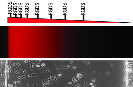
Microscale technologies are emerging as powerful tools for tissue engineering and biological studies. In this review, we present an overview of these technologies in various tissue engineering applications, such as for fabricating 3D microfabricated scaffolds, as templates for cell aggregate formation, or for fabricating materials in a spatially regulated manner. In addition, we give examples of the use of microscale technologies for controlling the cellular microenvironment in vitro and for performing high-throughput assays. The use of microfluidics, surface patterning, and patterned cocultures in regulating various aspects of cellular microenvironment is discussed, as well as the application of these technologies in directing cell fate and elucidating the underlying biology. Throughout this review, we will use specific examples where available and will provide trends and future directions in the field.


Conflict of interest statement

Conflict of interest statement: No conflicts declared.

Figures

Fig. 1.
Fig. 1.
Tissue engineering approaches. Tissue engineering approaches are classified into three categories: (i) cells alone, (ii) cells with scaffolds, and (iii) scaffolds alone. Each one of these approaches can be enhanced by in vitro microenvironmental factors before application as a tissue substitute.
Fig. 2.
Fig. 2.
Microscale technologies for tissue engineering. (A) Microtechnologies can be used directly to fabricate improved scaffolds and bioreactors or indirectly to study cellular behavior in controlled conditions or through the use of high-throughput experimentation. (B) A PDMS microfabricated tissue engineering scaffold with the vasculature directly embedded into the scaffold (17). (C) Various microscale techniques used to control different aspects of cell–microenvironment interactions are shown.
Fig. 3.
Fig. 3.
Gradient hydrogels for tissue engineering. (Top) Hydrogels can be fabricated with control over the spatial properties of the materials by embedding a gradient of materials, such as RGD peptide, directly into the material. (Middle and Bottom) The shape of the gradient can be visualized by using fluorescent molecules (Middle), and its function can observed by imaging endothelial cell adhesion after a few hours on the gels (Bottom) (39).
Fig. 4.
Fig. 4.
Microscale tissue engineering using template-based cell assembly. (A) A schematic diagram of the template-based assembly method. PEG microwells were fabricated so that cells could dock within the low-shear-stress regions generated within the microstructures. Once cells had immobilized within the microwells, other cells were washed away, and the cells within the microwells formed aggregates of controlled properties. (B) A light microscope image of 100-μm PEG wells that were seeded with ES cells and washed. (C) A scanning electron micrograph of cells within PEG microwells (A.K., J. Yeh, G. Eng, J. Fukuda, O. Farokhzad, J. J. Cheng, J. Bumbling, and R.L., unpublished data).
Fig. 5.
Fig. 5.
Microscale approaches for controlling the in vitro cellular microenvironment. (A) Light microscope images of ES cells patterned on PEG-coated substrates as an example of surface patterning for regulating cell–ECM interactions. (B) A fluorescent image of patterned cocultures depicting control over the degree of heterotypic and homotypic interactions between ES cells and fibroblasts (95). (C) An image of a cell and a schematic diagram of microfluidic methods of regulating cell-soluble signal interactions by flowing two parallel streams of fluids on an individual cell (81).
Fig. 6.
Fig. 6.
Stem cell arrays for tracking cell fates in culture. (A) Cell microarrays used to track clonal populations of stem cells after 4 days. Green boxes had proliferating cells, whereas red boxes did not contain proliferating cells (76). (B) Fluorescent images of the differentiating cells inside a microfabricated array that were stained with Tuj1 (red) and glial fibrillary acidic protein (blue) (76). (Scale bar, 100 μm.)
Fig. 7.
Fig. 7.
Light microscope images of human mesenchymal stem cells on small and large fibronectin islands after 1 week of culture. The images indicate that cells on the small islands stained for lipids, thus differentiating into adipogenic fates, whereas cells on large islands stained for alkaline phosphatase and differentiated into osteoblasts (78). (Scale bars, 50 μm.)
Fig. 8.
Fig. 8.
Schematic diagram of the procedure used to fabricate arrays of polymeric biomaterials (103).

References

    1. Langer R., Vacanti J. P. Science. 1993;260:920–926. - PubMed
    1. Griffith L. G., Naughton G. Science. 2002;295:1009–1014. - PubMed
    1. Niklason L. E., Langer R. J. Am. Med. Assoc. 2001;285:573–576. - PubMed
    1. Andersson H., van den Berg A. Lab Chip. 2004;4:98–103. - PubMed
    1. Whitesides G. M., Ostuni E., Takayama S., Jiang X. Y., Ingber D. E. Annu. Rev. Biomed. Eng. 2001;3:335–373. - PubMed

Publication types

LinkOut - more resources