. 2006 Feb 21;103(8):2827-32.
doi: 10.1073/pnas.0510712103. Epub 2006 Feb 13.

The U(L)41 protein of herpes simplex virus 1 degrades RNA by endonucleolytic cleavage in absence of other cellular or viral proteins

Affiliations

The U(L)41 protein of herpes simplex virus 1 degrades RNA by endonucleolytic cleavage in absence of other cellular or viral proteins

Brunella Taddeo et al. Proc Natl Acad Sci U S A. .

Abstract

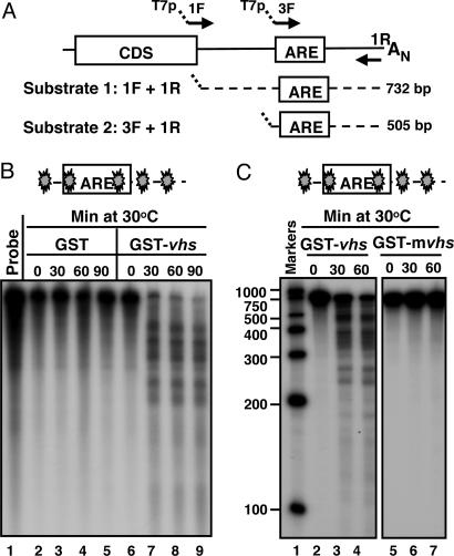
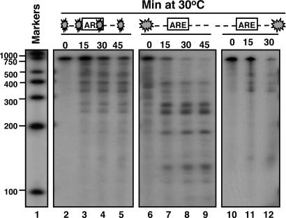
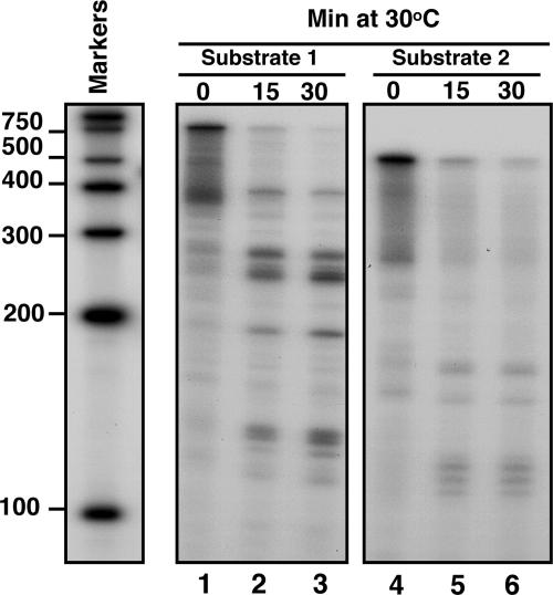
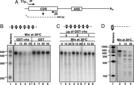
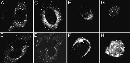
The herpes simplex virus 1 ORF U(L)41 encodes a protein (virion host shutoff or vhs) associated with selective degradation of mRNA early in infection. Some mRNAs, exemplified by GAPDH or beta-actin mRNAs, are degraded rapidly. Others, for example IEX-1 mRNA, are degraded in two stages: whereas the 3' domain disappears rapidly, a large 5' domain fragment of the mRNA lingers for several hours. Still a third, exemplified by tristetraprolin mRNA, is not degraded, allowing its protein product to accumulate in infected cells. Here we report the following: (i) a GST-vhs protein produced in Escherichia coli, solubilized and purified to homogeneity acts as bona fide endoribonuclease when tested on in vitro transcribed IEX-1 probes. A GST-vhs protein in which three key vhs amino acids were replaced with alanines, solubilized and purified by the same protocol, had no enzymatic activity. (ii) The number of fragments generated by cleavage of a truncated IEX-1 RNA by vhs appears to be small; the cleavage sites are centered at or near the AU-rich elements located at the 3' untranslated region of the mRNA. A truncated RNA containing only the IEX-1 coding domain was cleaved numerous times. (iii) In cells infected at high multiplicity and exposed to a large number of particles per cell, the vhs protein accumulated within 3 h after infection, in small uniform cytoplasmic granules raising the possibility that vhs colocalizes with tristerapolin, a protein induced after infection, in structures involved in degradation of RNA.


Conflict of interest statement

Conflict of interest statement: No conflicts declared.

Figures

Fig. 1.
Fig. 1.
Expression of recombinant GST-vhs fusion protein and specificity of anti-vhs antiserum. (A) Coomassie blue-stained gel of GST-vhs fusion protein bound to GS and eluted with buffer containing 75 mM Hepes (pH 7.4), 150 mM NaCl, 5 mM DTT, and 0.08% SDS before (lane 2) and after (lane 3) dialysis against buffer lacking SDS. The molecular weight marker is also shown (lane 1). (B) HeLa and Hep2 cells were either mock infected (lane 1) or infected with 10 PFU/cell of HSV-1(F) (lane 2), ΔUL41 mutant virus (lane 3) or ΔUL41R repaired virus (lane 4). Cells were harvested 18 h after infection and subjected to electrophoretic separation in denaturing gels and immunoblotting with the anti-vhs rabbit antiserum.
Fig. 2.
Fig. 2.
GST-vhs fusion protein is biologically active. (A) Schematic representation of the full-length IEX-1 mRNA and location of the primers used to generate the DNA templates for in vitro transcription of substrates 1 and 2. (B) Five micrograms of either GST (lanes 2–5) or GST-vhs fusion protein (lanes 6–9) were incubated at 30°C with 32P internally labeled IEX-1 3′ UTR RNA (substrate 1). Samples were removed at the indicated times, purified, and resolved on a sequencing gel, followed by autoradiography as described in Materials and Methods. Free probe (lane 1) was extracted and analyzed as well. (C) Five micrograms of either GST-vhs wild-type protein (lanes 2–4) or GST-mvhs mutant protein, bearing alanine substitutions of E192, D194, and D195 (lanes 5–7) were incubated at 30°C with 32P internally labeled substrate 1 for time intervals shown and RNA samples were analyzed as in A. Internally labeled RNA marker was loaded in lane 1, and the length of the fragments is reported on the side. The filled symbol marks the site of labeling of the RNA.
Fig. 3.
Fig. 3.
vhs cleavage sites in truncated transcripts containing AREs. Substrate 1 was either 32P internally labeled (lanes 2–5), or labeled at the 5′end (lanes 6–9) or the 3′end (lanes 10–12). The substrates were incubated at 30°C with 5 μg of purified GST-vhs for the indicated times. Samples were purified and resolved on a sequencing gel, followed by autoradiography as described in Materials and Methods. Internally labeled RNA marker was loaded in lane 1, and the length of the fragments is reported on the side.
Fig. 4.
Fig. 4.
vhs cleaves truncated transcripts containing AREs at preferential sites. Substrates 1 (lanes 1–3) and 2 (lanes 4–6) were labeled with γ32P at the 5′end and incubated with 5 μg of purified GST-vhs protein at 30°C for the indicated times. Samples were purified and resolved on a sequencing gel, followed by autoradiography as described in Materials and Methods. Internally labeled RNA marker was also loaded, and the length of the fragments is reported on the side.
Fig. 5.
Fig. 5.
vhs cleaves truncated transcripts containing the IEX-1 coding domains at multiple sites. (A) Schematic representation of the full-length IEX-1 mRNA and location of the primers used to generate the DNA template for in vitro transcription of IEX-1 coding domain (substrate 3). (B) Five micrograms of either GST-vhs fusion protein (lanes 2–5) or GST (lanes 6–9) were incubated at 30°C with internally labeled substrate 3. Samples were removed at the indicated times, purified, and resolved on a sequencing gel, followed by autoradiography as described in Materials and Methods. Internally labeled RNA marker was loaded in lane 1, and the length of the fragments is reported on the side. (C) Internally labeled substrate 3 was incubated with different amounts of GST-vhs fusion protein: 4 μg (lanes 1–3), 2 μg (lanes 4–6), and 1 μg (lanes 7–9). Samples were analyzed as in B. (D) 5′ end-labeled substrate 3 was incubated with 5 μg of purified GST-vhs fusion protein at 30°C for the indicated times. Samples were analyzed as in B and C.
Fig. 6.
Fig. 6.
vhs introduced into cells during infection and newly made vhs accumulate in the cytoplasm and perinuclear region of infected cells. (AD) Representative SK-N-SH cells exposed to 100 PFU of purified HSV-1(F) and fixed and stained with primary anti-vhs and secondary anti-rabbit conjugated antibody at 3 h after infection. (EH) HEp-2 cells fixed 16 h after exposure to 10 PFU of HSV-1(F) and reacted with either anti-vhs (E and F) or anti-VP22 antibodies (G and H). The cultures were reacted with secondary antibody conjugated with fluorescein isothiocyanate as described in Materials and Methods.

References

    1. Roizman B., Borman G. S., Kamali-Rousta M. Nature. 1965;206:1374–1375. - PubMed
    1. Kwong A. D., Frenkel N. Proc. Natl. Acad. Sci. USA. 1987;84:1926–1930. - PMC - PubMed
    1. Strom T., Frenkel N. J. Virol. 1987;61:2198–2207. - PMC - PubMed
    1. Karr B. M., Read G. S. Virology. 1999;264:195–204. - PubMed
    1. Kwong A. D., Kruper J. A., Frenkel N. J. Virol. 1988;62:912–921. - PMC - PubMed

Publication types

MeSH terms