. 2006 Feb 15;81(3):438-44.
doi: 10.1097/01.tp.0000188948.72706.4d.

The difficulty of eliminating donor leukocyte microchimerism in rat recipients bearing established organ allografts

Affiliations

The difficulty of eliminating donor leukocyte microchimerism in rat recipients bearing established organ allografts

Tetsuma Kiyomoto et al. Transplantation. .

Abstract

Background: Unequivocal eradication of donor leukocyte microchimerism from recipients of long-surviving organ transplants has never been reported. Here we describe a drastic attempt to accomplish this objective.

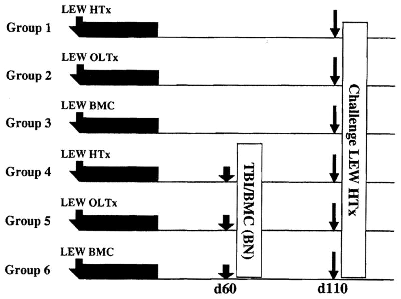
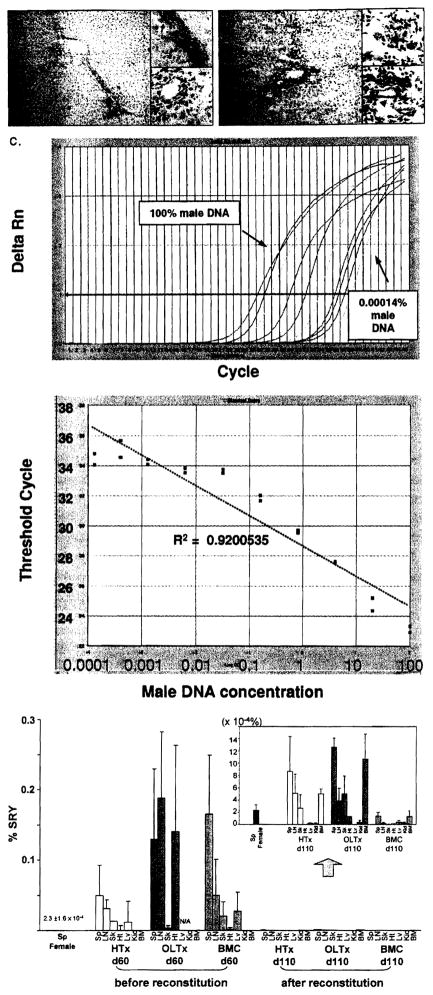
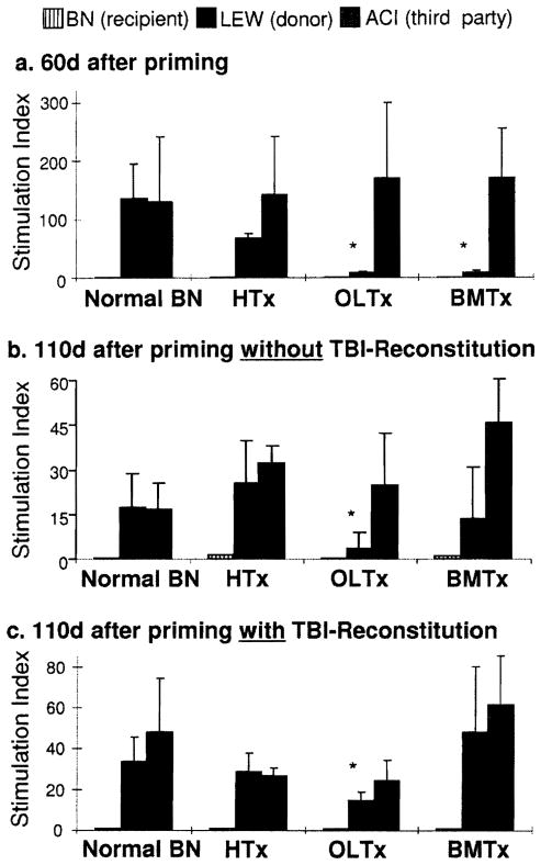
Methods: In control experiments, a rank order of microchimerism and of associated donor specific nonreactivity was produced in Brown-Norway (BN) rats by transplantation of Lewis (LEW) liver, bone marrow cell (BMC) and heart allografts under a brief course of tacrolimus. The degree of microchimerism at 60 and 110 days was estimated with semiquanitative immunocytochemical and PCR techniques. Tolerance at 110 days was assessed in the different control groups by challenge transplantation of naïve LEW hearts. In parallel experimental groups, an attempt was made to eliminate microchimerism from the BN recipients. The animals were submitted at 60 days to 9.5-Gy total body irradiation (TBI), reconstituted immediately with naïve BN BMC, and tested for donor specific nonreactivity by LEW heart transplantation at 110 days.

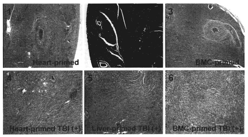
Results: After the TBI-reconstitution at 60 days, microchimerism was undetectable in BMC recipients at 110 days, significantly reduced in heart recipients, and least affected in liver recipients. Except in liver recipients, abrogation of LEW-specific nonreactivity was demonstrated by rejection of the priming grafts, or by rejection of the challenge heart grafts, and by in vitro immune assay.

Conclusions: It is difficult to eliminate microchimerism in organ recipients once the donor cells have settled into tissue niches.


Figures

FIGURE 1
FIGURE 1
Experimental design in which all six groups of BN recipients were given LEW priming allografts under a short course of tacrolimus (TAC) immunosuppression and tested for donor-specific reactivity 110 days later by challenge LEW heart transplantation (bold arrows). No other treatment was given to the animals of groups 1–3. In Groups 4–6, it was attempted to remove donor leukocyte micro-chimerism by 9.5 Gy TBI and immediate reconstitution with 5×10⁷ naïve BN BMC (shaded arrows). Transplantations were of heterotopic hearts (HTx), orthotopic livers (OLTx), and infused bone marrow cells (BMC).
FIGURE 2
FIGURE 2
Microchimerism after 110 days in female BN recipients primed with male LEW allografts before and after TBI reconstitution. (A) Transplanted LEW liver after 110 days in non-irradiated BN recipients (see Group 2, Fig. 1). LEW MHC class II+ cells in the periportal area are stained brown by the L21–6 mAb (arrows, insert, upper). Note that biliary epithelial cells remain negative for L21–6 (insert, lower). Original magnification, X200 and X600. (B) Transplanted LEW liver after 110 days in irradiated and reconstituted BN recipients (see Group 5, Fig. 1). Most of the donor MHC class II+ cells are missing, but some remain (arrows, insert, upper). Faint staining of biliary epithelial cells (arrow heads, insert, lower) suggests the possibility of subtle biliary injury that was not detectable with conventional histopathology. Three animals in each group were analyzed and representative picture is shown. Original magnification, X200 and X600. (C) Standardized amplification curve made with artificial mixtures of serial male DNA concentrations. Artificial mixtures were made with serial male DNA concentrations (10 serial dilutions of 100, 20, 4, 0.8, 0.16, 0.032, 0.0064, 0.00128, 0.000427, 0.000142% male DNA) and analyzed with SYB1 green real-time PCR. Upper panel shows Sry-specific marker ΔRn curves for the 10 samples. Regular positive amplification curves were observed with ΔRn curve shift to the right as male DNA concentration decreased. Lowe panel shows standardized amplification curve plotted from these results. Cycle threshold values linearly correlated with the log of male DNA concentration (R2=0.9200535). (D) Male DNA concentrations (microchimerism) before (60 days) and after (110 days) TBI-reconstitution in tissues of female recipients of male heart, liver, and BMC allografts (Groups 4–6 in Figure 1, n=3 in each with SYBR green real-time PCR). At 60 days before TBI-reconstitution, ail primed recipients with LEW heart, liver and BMC showed ~0.1 % donor male DNA. After TBI-reconstitution, the male DNA found in liver recipients (~0.001%) was present in smaller amounts in heart recipients, but was not identifiable in BMC recipients (insert). BM, bone marrow; LN, lymph nodes; Sp, spleen; Kid, kidney; Sk, skin; Ht, heart; Lv, liver.
FIGURE 3
FIGURE 3
One-way MLR of BN cells (from cervical nodes) to irradiated stimulator cells. (A) 60 days: before TBI/reconstitution. (B) 110 days: without TBI/reconstitution. (C) 110 days: after TBI/reconstitution at 60 days. All results are expressed as mean stimulation index ± SD n=3 each group. *P<0.05 vs. normal BN response to LEW stimulator cells. HTx, heart transplantation; OLTx, orthotopic liver transplantation; BMTx, bone marrow cell transplantation.
FIGURE 4
FIGURE 4
Histopathological findings of challenge heart allografts after 100 days (210 days after primary transplantation). The six panel numbers correspond to the six groups shown in Figure 1 and histological findings in Table 1. H&E stain, original magnification × 100.

References

    1. Starzl TE, Demetris AJ, Murase N, et al. Cell migration, chimerism, and graft acceptance. Lancet. 1992;339(8809):1579. - PMC - PubMed
    1. Nemlander A, Soots A, von Willebrand E, et al. Redistribution of renal allograft-responding leukocytes during rejection. II. Kinetics and specificity. J Exp Med. 1982;156:1087–1100. - PMC - PubMed
    1. Larsen CP, Morris PJ, Austyn JM. Migration of dendritic leukocytes from cardiac allografts into host spleens. A novel route for initiation of rejection. J Exp Med. 1990;171:307–314. - PMC - PubMed
    1. Demetris AJ, Qian S, Sun H, et al. Early events in liver allograft rejection. Am J Pathol. 1991;138:609–618. - PMC - PubMed
    1. Demetris AJ, Murase N, Fujisaki S, et al. Hematolymphoid cell trafficking, microchimerism, and GVHD reactions after liver, bone marrow, and heart transplantation. Transplant Proc. 1993;25:3337–3344. - PMC - PubMed

MeSH terms