

Developmental expression patterns of Tbx1, Tbx2, Tbx5, and Tbx20 in Xenopus tropicalis
- PMID: 16477648
- PMCID: PMC1635807
- DOI: 10.1002/dvdy.20714
Developmental expression patterns of Tbx1, Tbx2, Tbx5, and Tbx20 in Xenopus tropicalis
Abstract
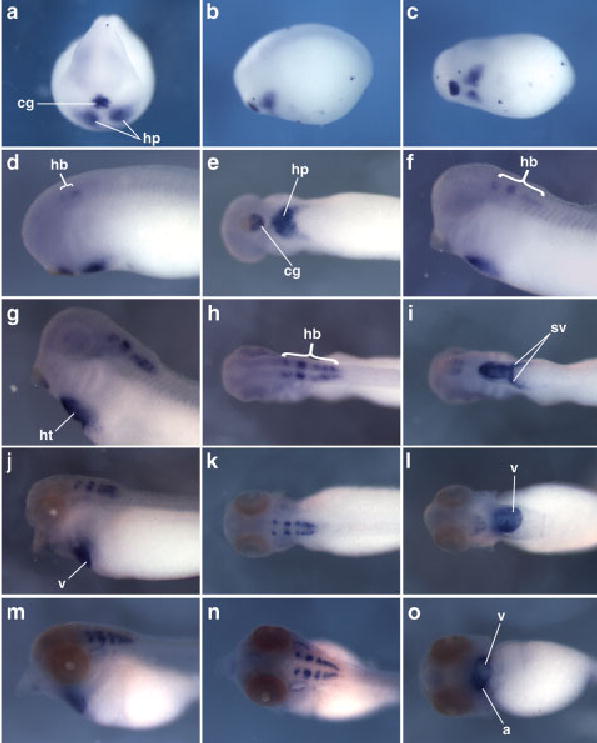
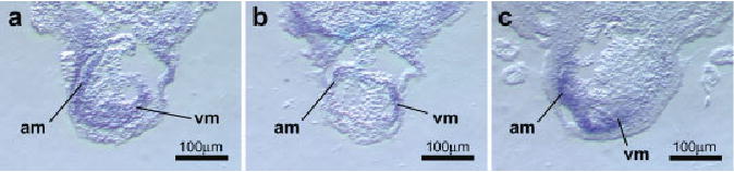
T-box genes have diverse functions during embryogenesis and are implicated in several human congenital disorders. Here, we report the identification, sequence analysis, and developmental expression patterns of four members of the T-box gene family in the diploid frog Xenopus tropicalis. These four genes-Tbx1, Tbx2, Tbx5, and Tbx20-have been shown to influence cardiac development in a variety of organisms, in addition to their individual roles in regulating other aspects of embryonic development. Our results highlight the high degree of evolutionary conservation between orthologs of these genes in X. tropicalis and other vertebrates, both at the molecular level and in their developmental expression patterns, and also identify novel features of their expression. Thus, X. tropicalis represents a potentially valuable vertebrate model in which to further investigate the functions of these genes through genetic approaches.
Developmental Dynamics 235:1623-1630, 2006. (c) 2006 Wiley-Liss, Inc.
Figures
Similar articles
-
Tbx5 and Tbx20 act synergistically to control vertebrate heart morphogenesis.Development. 2005 Feb;132(3):553-63. doi: 10.1242/dev.01596. Epub 2005 Jan 5. Development. 2005. PMID: 15634698 Free PMC article.
-
Developmental expression of the N-myc downstream regulated gene (Ndrg) family during Xenopus tropicalis embryogenesis.Int J Dev Biol. 2015;59(10-12):511-7. doi: 10.1387/ijdb.150178xh. Int J Dev Biol. 2015. PMID: 26864492
-
The Xenopus Bowline/Ripply family proteins negatively regulate the transcriptional activity of T-box transcription factors.Int J Dev Biol. 2009;53(4):631-9. doi: 10.1387/ijdb.082823kh. Int J Dev Biol. 2009. PMID: 19247927
-
The Roles of T-Box Genes in Vertebrate Limb Development.Curr Top Dev Biol. 2017;122:355-381. doi: 10.1016/bs.ctdb.2016.08.009. Epub 2016 Oct 5. Curr Top Dev Biol. 2017. PMID: 28057270 Review.
-
Targeted integration of genes in Xenopus tropicalis.Genesis. 2017 Jan;55(1-2). doi: 10.1002/dvg.23006. Genesis. 2017. PMID: 28095621 Review.
Cited by
-
Tbx2 is required for the suppression of mesendoderm during early Xenopus development.Dev Dyn. 2018 Jul;247(7):903-913. doi: 10.1002/dvdy.24633. Epub 2018 May 4. Dev Dyn. 2018. PMID: 29633424 Free PMC article.
-
Tbx2a is required for specification of endodermal pouches during development of the pharyngeal arches.PLoS One. 2013 Oct 10;8(10):e77171. doi: 10.1371/journal.pone.0077171. eCollection 2013. PLoS One. 2013. PMID: 24130849 Free PMC article.
-
RIPPLY3 is a retinoic acid-inducible repressor required for setting the borders of the pre-placodal ectoderm.Development. 2012 Mar;139(6):1213-24. doi: 10.1242/dev.071456. Development. 2012. PMID: 22354841 Free PMC article.
-
Neuromancer1 and Neuromancer2 regulate cell fate specification in the developing embryonic CNS of Drosophila melanogaster.Dev Biol. 2009 Jan 1;325(1):138-50. doi: 10.1016/j.ydbio.2008.10.006. Epub 2008 Nov 1. Dev Biol. 2009. PMID: 19013145 Free PMC article.
-
How insights from cardiovascular developmental biology have impacted the care of infants and children with congenital heart disease.Mech Dev. 2012 Jul;129(5-8):75-97. doi: 10.1016/j.mod.2012.05.005. Epub 2012 May 26. Mech Dev. 2012. PMID: 22640994 Free PMC article. Review.
References
-
- Ahn DG, Ruvinsky I, Oates AC, Silver LM, Ho RK. tbx20, a new vertebrate T-box gene expressed in the cranial motor neurons and developing cardiovascular structures in zebrafish. Mech Dev. 2000;95:253–258. - PubMed
-
- Altschul SF, Gish W, Miller W, Myers EW, Lipman DJ. Basic local alignment search tool. J Mol Biol. 1990;215:403–410. - PubMed
-
- Ataliotis P, Ivins S, Mohun TJ, Scambler PJ. XTbx1 is a transcriptional activator involved in head and pharyngeal arch development in Xenopus laevis. Dev Dyn. 2005;232:979 –991. - PubMed
-
- Basson CT, Bachinsky DR, Lin RC, Levi T, Elkins JA, Soults J, Grayzel D, Kroumpouzou E, Traill TA, Leblanc-Straceski J, Renault B, Kucherlapati R, Seidman JG, Seidman CE. Mutations in human TBX5 [corrected] cause limb and cardiac malformation in Holt-Oram syndrome. Nat Genet. 1997;15:30 –35. - PubMed
Publication types
MeSH terms
Substances
Grants and funding
LinkOut - more resources
Full Text Sources
