Comparative Study
. 2006 Apr;83(5):845-56.
doi: 10.1002/jnr.20778.

Regulation of human neural precursor cells by laminin and integrins

Affiliations
Comparative Study

Regulation of human neural precursor cells by laminin and integrins

Lisa A Flanagan et al. J Neurosci Res. 2006 Apr.

Abstract

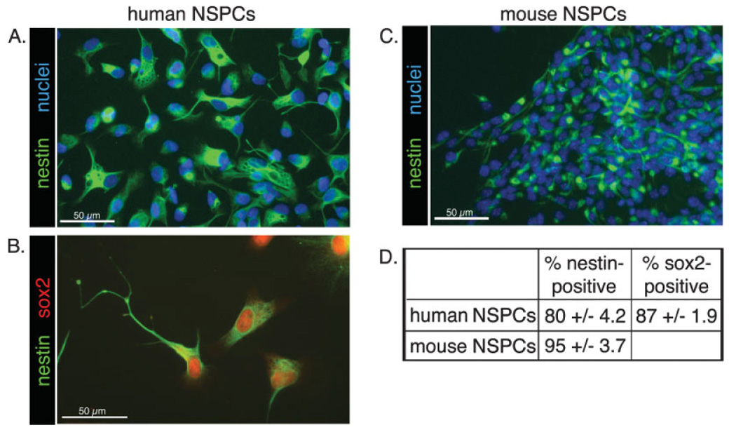
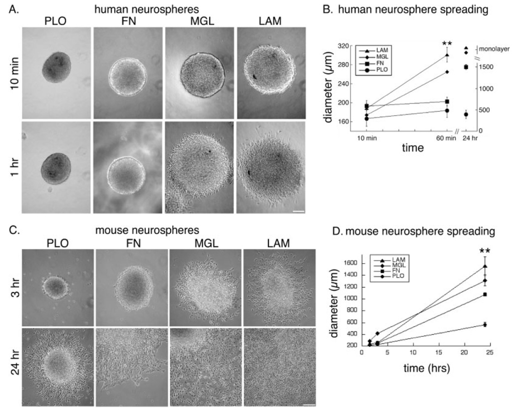
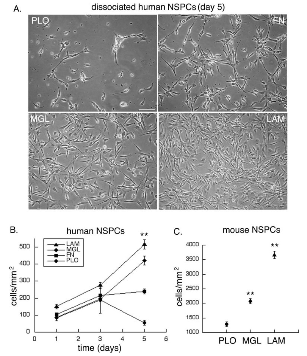
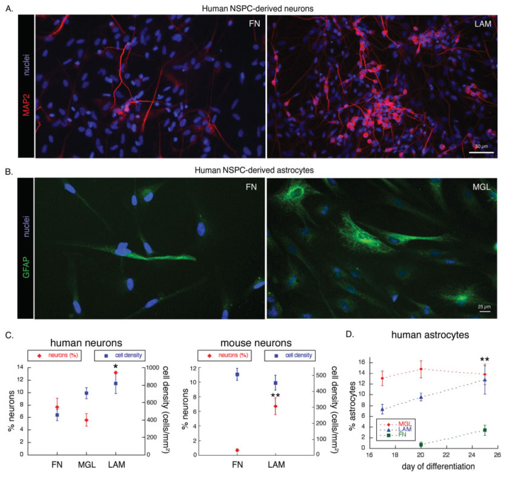
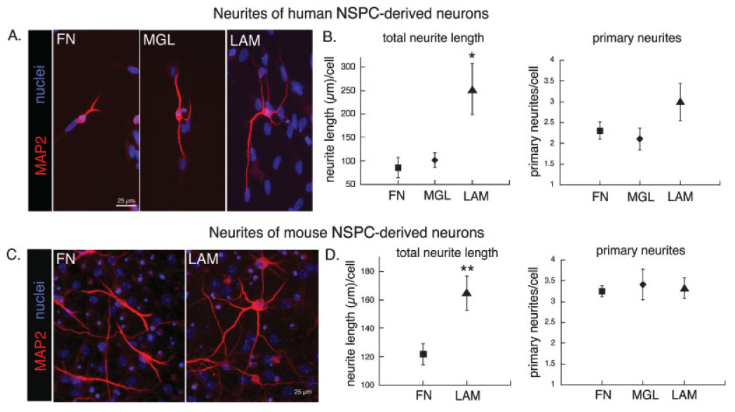
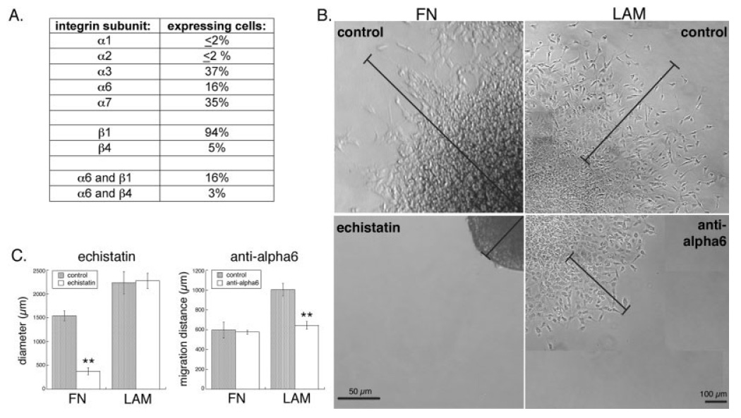
Deciphering the factors that regulate human neural stem cells will greatly aid in their use as models of development and as therapeutic agents. The extracellular matrix (ECM) is a component of stem cell niches in vivo and regulates multiple functions in diverse cell types, yet little is known about its effects on human neural stem/precursor cells (NSPCs). We therefore plated human NSPCs on four different substrates (poly-L-ornithine, fibronectin, laminin, and matrigel) and compared their responses with those of mouse NSPCs. Compared with the other substrates, laminin matrices enhanced NSPC migration, expansion, differentiation into neurons and astrocytes, and elongation of neurites from NSPC-derived neurons. Laminin had a similar spectrum of effects on both human and mouse cells, highlighting the evolutionary conservation of NSPC regulation by this component of the ECM. Flow cytometry revealed that human NSPCs express on their cell surfaces the laminin-binding integrins alpha3, alpha6, alpha7, beta1, and beta4, and function-blocking antibodies to the alpha6 subunit confirmed a role for integrins in laminin-dependent migration of human NSPCs. These results define laminin and its integrin receptors as key regulators of human NSPCs.


Figures

Fig. 1
Fig. 1
Most cultured human and mouse cortical cells express NSPC markers. A–C: Cultured cortical cells were immunostained to detect the NSPC markers nestin and sox2 (B). Nuclei were counterstained with Hoechst (A,C). D: Positively stained cells were expressed as a percentage of the total number of cells as determined by Hoechst-stained nuclei ± SEM.
Fig. 2
Fig. 2
Migration of human and mouse cortical NSPCs out of neurospheres is enhanced on laminin-containing matrices. A: Human neurospheres (SC23) were imaged at 10 min and 1 hr after plating onto the different substrates (PLO, poly-L-ornithine; FN, fibronectin; MGL, matrigel; LAM, laminin). B: Migration of human NSPCs (SC23 and SC27) was quantified by measuring the diameter of the sphere and surrounding rim of migrating cells over time. Human NSPCs on matrigel and laminin formed a dense monolayer by 24 hr that prevented accurate quantitation of migration (**P < 0.01 for diameter on LAM vs. FN or PLO; error bars represent SEM). C: Mouse neurospheres were imaged at 3 hr and 24 hr after plating (neurospheres are located in the upper left corners of the 24-hr images of cells on FN, MGL, and LAM to show the extent of cellular migration from the sphere). D: Quantitation of mouse NSPC migration over a 24-hr period was performed as described for human NSPCs (**P < 0.01 for diameter on LAM or MGL vs. FN or PLO and for FN vs. PLO; error bars represent SEM). Scale bars = 100 µm.
Fig. 3
Fig. 3
Expansion of human and mouse NSPCs is greater on laminin-containing matrices. Equal numbers of dissociated NSPCs were plated on substrate-coated coverslips, then cultured for varying lengths of time. A: Images of human NSPCs (SC23) cultured for 5 days on the different substrates. B: Total number of human NSPCs per surface area was counted on the different substrates over time (**P < 0.01 for cells/mm2 on LAM, MGL, or FN vs. PLO and for LAM or MGL vs. FN; P < 0.05 for LAM vs. MGL; error bars represent SEM). C: Mouse NSPC numbers were determined after culturing the cells on the different substrates for 3 days (**P < 0.01 for cells/mm2 on LAM or MGL vs. PLO and for LAM vs. MGL; error bars represent SEM). Scale bar = 100 µm.
Fig. 4
Fig. 4
Greater numbers of neurons and astrocytes are derived from human NSPCs on laminin. A: Human NSPCs (SC27) were differentiated for 20 days on FN or LAM and immunostained to detect expression of the neuronal marker MAP2. All nuclei were stained by Hoechst. Similar patterns of expression of neuronal markers were obtained with two other human NSPC isolates (SC23 and SC30; data not shown). B: Human NSPCs (SC23) were differentiated for 20 days on FN or MGL and astrocytes visualized by immunostaining for GFAP. C: Human NSPCs were differentiated for 20 days on the various substrates, and cells expressing the neuronal marker MAP2 were counted and expressed as a percentage of the total number of cells (determined by Hoechst stained nuclei; *P < 0.05 for percent neurons on LAM vs. FN and P < 0.01 for percentage neurons on LAM vs. MGL; error bars represent SEM). The percentage of neurons (red diamonds) and total cell density (blue squares) are graphed together to illustrate that the effect of substrate on neuronal number is independent of cell density. Mouse NSPCs were differentiated for 10 days on the various substrates, and cells expressing neuronal markers (either MAP2 or TuJ1) were counted and expressed as a percentage of the total number of cells (red diamonds; **P < 0.01 for percent neurons on LAM vs. FN; error bars represent SEM). The density of mouse NSPCs did not differ on LAM vs. FN (blue squares). D: Numbers of GFAP-positive astrocytes were counted after 17–25 days of differentiation on the various substrates and expressed as a percentage of the total number of cells (**P < 0.01 for percent astrocytes on LAM or MGL vs. FN; error bars represent SEM).
Fig. 5
Fig. 5
Neurons generated from human and mouse NSPCs extend longer neurites on laminin. Human or mouse NSPCs were differentiated on the various substrates to allow formation of neurons with concomitant extension of neurites. A: Human NSPCs (SC23) were differentiated for 25 days and neurites visualized by immunostaining for MAP2. Nuclei were counterstained with Hoechst. B: Total neurite length and number of primary neurites were determined for each human NSPC-derived neuron (see Materials and Methods for details of quantification; *P < 0.05 for total neurite length on LAM vs. MGL or FN; error bars represent SEM). No difference was found in the number of neurite branches (see text). C: Mouse NSPCs were differentiated for 10 days on the various substrates and immunostained for MAP2 to visualize neurites (nuclei counterstained with Hoechst). D: Total neurite length and numbers of primary neurites were quantified for mouse NSPC-derived neurons as for human neurons (**P < 0.01 for total neurite length on LAM vs. FN; error bars represent SEM). As for human cells, there was no difference in neurite branching (see text).
Fig. 6
Fig. 6
Cell surface integrins mediate responses of human NSPCs to extracellular matrices. A: Human NSPCs (SC27) were analyzed by flow cytometry to determine the mean percentages of cells expressing the laminin-binding α and β integrin subunits and the percentage of cells that coexpress the α6 subunit with its binding partners β1 and β4. Similar levels of integrin subunit expression were detected on SC30 human NSPCs. B: Human NSPC spheres (SC27) were adhered to FN- or LAM-coated coverslips, then treated with the disintegrin echistatin (FN panels) or an α6 integrin blocking antibody (LAM panels). Bars in the FN panels denote the distance from the middle of the sphere to the edge of migrating cells at 24 hr (control) or to the edge of the nonadherent sphere (echistatin). Bars in the LAM panels indicate the extent of NSPC migration by marking the position of the front of migrating cells prior to antibody addition and after 24 hr. C: Quantitation of spheres and migrating cells at 24 hr shows that human NSPCs migrate out of control spheres on FN, but spheres treated with echistatin were no longer adhered and thus cells were unable to migrate (**P < 0.01). Migration on LAM was partially blocked by an α6 antibody (see Materials and Methods for description of quantitation). Controls in B and C are no treatment (for echistatin) or incubation with an isotype-matched control antibody (for anti-α6).

References

    1. Alvarez-Buylla A, Lim DA. For the long run: maintaining germinal niches in the adult brain. Neuron. 2004;41:683–686. - PubMed
    1. Anlar B, Atilla P, Cakar AN, Kose MF, Beksac MS, Dagdeviren A, Akcoren Z. Expression of adhesion and extracellular matrix molecules in the developing human brain. J Child Neurol. 2002;17:707–713. - PubMed
    1. Bokel C, Brown NH. Integrins in development: moving on, responding to, and sticking to the extracellular matrix. Dev Cell. 2002;3:311–321. - PubMed
    1. Burgeson RE, Chiquet M, Deutzmann R, Ekblom P, Engel J, Kleinman H, Martin GR, Meneguzzi G, Paulsson M, Sanes J, Timpl R, Tryggvason K, Yamada Y, Yurchenco PD. A new nomenclature for the laminins. Matrix Biol. 1994;14:209–211. - PubMed
    1. Cai J, Wu Y, Mirua T, Pierce JL, Lucero MT, Albertine KH, Spangrude GJ, Rao MS. Properties of a fetal multipotent neural stem cell (NEP cell) Dev Biol. 2002;251:221–240. - PubMed

Publication types

MeSH terms