. 2006 May 15;396(1):23-30.
doi: 10.1042/BJ20051568.

RanBPM associates with CD39 and modulates ecto-nucleotidase activity

Affiliations

RanBPM associates with CD39 and modulates ecto-nucleotidase activity

Yan Wu et al. Biochem J. .

Abstract

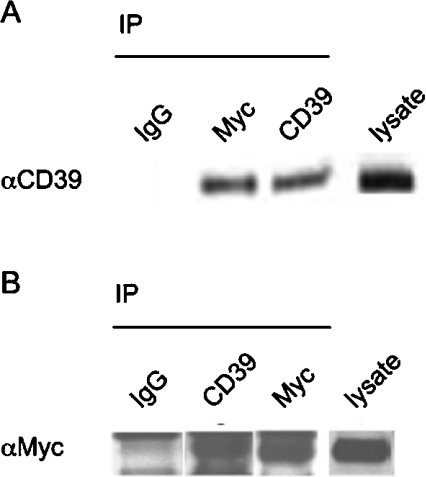
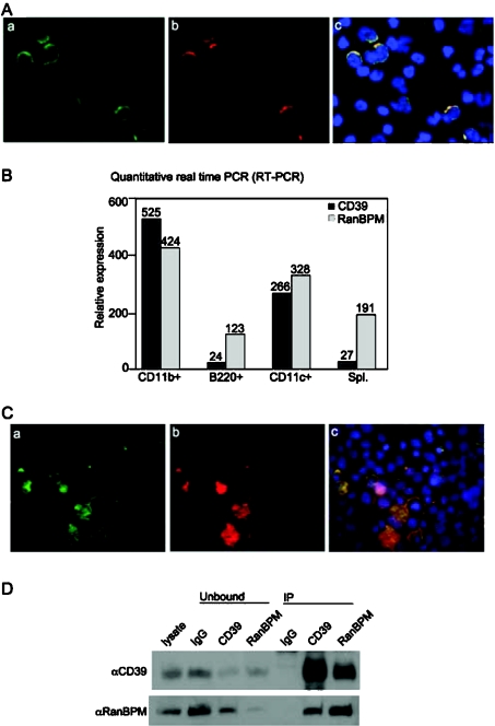
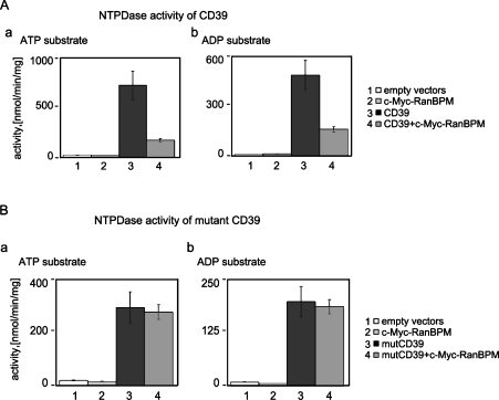
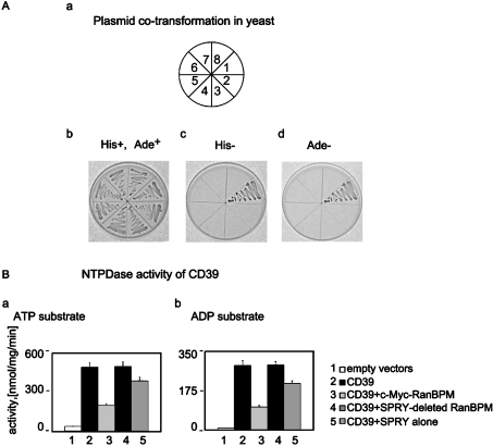
CD39/ecto-NTPDase 1 (nucleoside triphosphate diphosphohydrolase 1) is an ecto-nucleotidase that influences P2 receptor activation to regulate vascular and immune cell adhesion and signalling events pivotal in inflammation. Whether CD39 interacts with other membrane or cytoplasmic proteins has not been established to date. Using the yeast two-hybrid system, we note that the N-terminus of CD39 binds to RanBPM (Ran binding protein M; also known as RanBP9), a multi-adaptor scaffolding membrane protein originally characterized as a binding protein for the small GTPase Ran. We confirm formation of complexes between CD39 and RanBPM in transfected mammalian cells by co-immunoprecipitation studies. Endogenous CD39 and RanBPM are also found to be co-expressed and abundant in cell membranes of B-lymphocytes. NTPDase activity of recombinant CD39, but not of N-terminus-deleted-CD39 mutant, is substantially diminished by RanBPM co-expression in COS-7 cells. The conserved SPRY [repeats in splA and RyR (ryanodine receptor)] moiety of RanBPM is insufficient alone for complete physical and functional interactions with CD39. We conclude that CD39 associations with RanBPM have the potential to regulate NTPDase catalytic activity. This intermolecular interaction may have important implications for the regulation of extracellular nucleotide-mediated signalling.


Figures

Figure 1
Figure 1. Co-immunoprecipitation of recombinant CD39 with c-Myc-tagged RanBPM
(A, B) Cell lysates (500 μg) from COS-7 cells expressing recombinant CD39 protein and c-Myc–RanBPM were subjected to immunoprecipitation (IP) with CD39-specific antibody or c-Myc-specific antibody or control mouse isotype-matched IgG1, as indicated on the top of the panels. The antibody-captured proteins were analysed by Western blotting (antibody specificity is identified to the left of the panels). CD39 blots were analysed under non-reducing conditions. c-Myc–RanBPM blots were analysed under reducing conditions. The lane on the far right shows the appropriate positive control Western-blot analysis of total lysates. αCD39, anti-CD39; αMyc, anti-Myc.
Figure 2
Figure 2. Endogenous associations of CD39 and RanBPM
(A) Triple immunofluorescent analysis for the endogenous co-localization of CD39 (a, green) and RanBPM (b, red) in human peripheral blood mononuclear cells. CD39 staining was performed using FITC-labelled monoclonal anti-CD39 antibody. RanBPM staining was performed using monoclonal anti-RanBPM antibody followed by chicken anti-mouse IgG conjugated with Alexa Fluor® 594. Nuclear staining was performed using Hoechst 33258 (c, blue). Co-localization of the two proteins in the cell membranes of peripheral blood mononuclear cells was observed (c, yellow, overlay of a and b). (B) Quantitative real-time PCR analysis of CD39 and RanBPM mRNA co-expression in murine CD11b+ cells (macrophages), B220+ cells (B-lymphocytes), CD11c+ cells (DC) and total splenocytes (Spl.). These cells all co-expressed CD39 and RanBPM at mRNA level. (C) Triple immunofluorescent staining was performed in EBV-transformed human B-lymphocytes, as described above. Endogenous CD39 (a, green) co-localized with endogenous RanBPM (b, red) in the cell membranes of B-lymphocytes (c, yellow, overlay of a and b). (D) RanBPM interacts with CD39 in B-lymphocytes. EBV-transformed B-lymphocyte lysates were immunoprecipitated with the indicated antibodies. Immunoprecipitates (IP) were resolved on a 4–15% gradient SDS/PAGE, transferred on to a PVDF membrane, and probed with antibodies as indicated to the left of the panels. The far-left lane shows control Western-blot analysis of total lysates (10% input). The residual non-immunoprecipitated proteins within the lysates are indicated as ‘Unbound’ at the top of the panel. CD39 was analysed under non-reducing conditions; RanBPM was analysed under reducing conditions, as before.
Figure 3
Figure 3. RanBPM inhibits the NTPDase activity of CD39 by interacting with the N-terminus of CD39
(A) ATP and ADP (as indicated at the top of the panels) are used as substrates of the NTPDase activity in intact transfected COS-7 cells overexpressing CD39 and/or c-Myc–RanBPM. COS-7 cells transfected with empty vectors were used as a negative control. Samples are: bar 1, control reaction with COS-7 cells transfected with empty plasmid vectors; bar 2, cells overexpressing c-Myc–RanBPM; bar 3, cells with ectopic CD39; bar 4, cells expressing both CD39 and c-Myc–RanBPM proteins. Three independent experiments produced consistent results (S.D.=±7%). (B) The same as described in (A) except for NT-Δ-1–37 CD39 mutant (mutCD39) instead of native CD39.
Figure 4
Figure 4. SPRY domains of RanBPM and interactions with the N-termini of CD39
(A) Probing for SPRY–CD39 interactions by yeast two-hybrid assays. Yeast PJ69-4A cells were co-transformed with the CD39 N-terminal ‘bait’ (pGBDU-C1-CD39N1-14) and either the originally deleted clone lacking N-terminal amino acids 1–148 (pACT2-RanBPM149-729) (position 1), or the SPRY-truncated RanBPM expression plasmid (pGADT7-RanBPM351-729) (position 2) and the SPRY domain alone (pGADT7-RanBPM149-353) (position 3). All the remaining co-transformations served as controls. These were conducted with: the ‘bait-less’ empty vector pGBDU-C1 and the empty control AD vector pGADT7 (position 4), ‘bait’ and pGADT7 (position 5), empty pGBDU-C1 and pACT2-RanBPM149-729 (position 6), pGBDU-C1 and pGADT7-RanBPM351-729 (position 7), and finally pGBDU-C1 and pGADT7-RanBPM149-353 (position 8). His+ and Ade+ indicate uracil- and leucine-deficient plates. His plates lack uracil, leucine and histidine. Ade plates lack uracil, leucine and adenine. Plates with the co-transformed yeast cells were incubated at 30 °C for 3 days. (B) SPRY domains, in isolation, are not sufficient for full functional interaction between RanBPM and CD39. NTPDase activity assay with ATP and ADP as substrates (indicated at the top of the panels) in COS-7 cells with CD39 alone (bar 2); CD39 and intact RanBPM (bar 3); CD39 and SPRY-truncated RanBPM variant (bar 4); CD39 and RanBPM with isolated SPRY moiety (bar 5). As a control COS-7 cells transfected with empty vectors lacking the protein inserts were used (bar 1). Three independent experiments produced consistent results (variance=±7%).

References

    1. Maliszewski C. R., Delespesse G. J., Schoenborn M. A., Armitage R. J., Fanslow W. C., Nakajima T., Baker E., Sutherland G. R., Poindexter K., Birks C. The CD39 lymphoid cell activation antigen. Molecular cloning and structural characterization. J. Immunol. 1994;153:3574–3583. - PubMed
    1. Kaczmarek E., Koziak K., Sevigny J., Siegel J. B., Anrather J., Beaudoin A. R., Bach F. H., Robson S. C. Identification and characterization of CD39/vascular ATP diphosphohydrolase. J. Biol. Chem. 1996;271:33116–33122. - PubMed
    1. Wang T. F., Guidotti G. CD39 is an ecto-(Ca2+,Mg2+)-apyrase. J. Biol. Chem. 1996;271:9898–9901. - PubMed
    1. Enjyoji K., Sevigny J., Lin Y., Frenette P. S., Christie P. D., Esch J. S., II, Imai M., Edelberg J. M., Rayburn H., Lech M., et al. Targeted disruption of cd39/ATP diphosphohydrolase results in disordered hemostasis and thromboregulation. Nat. Med. 1999;5:1010–1017. - PubMed
    1. Goepfert C., Imai M., Brouard S., Csizmadia E., Kaczmarek E., Robson S. C. CD39 modulates endothelial cell activation and apoptosis. Mol. Med. 2000;6:591–603. - PMC - PubMed

Publication types

MeSH terms